

Why Are Dark Patches Showing Up on My Cheeks After an Eyelid Rash?
A 40-year-old woman presented to Village Dermatology in Katy, Texas with dark cheek discoloration following an eyelid rash. Learn about post-inflammatory hyperpigmentation treatment and expert dermatologic care in Houston and Katy, TX.
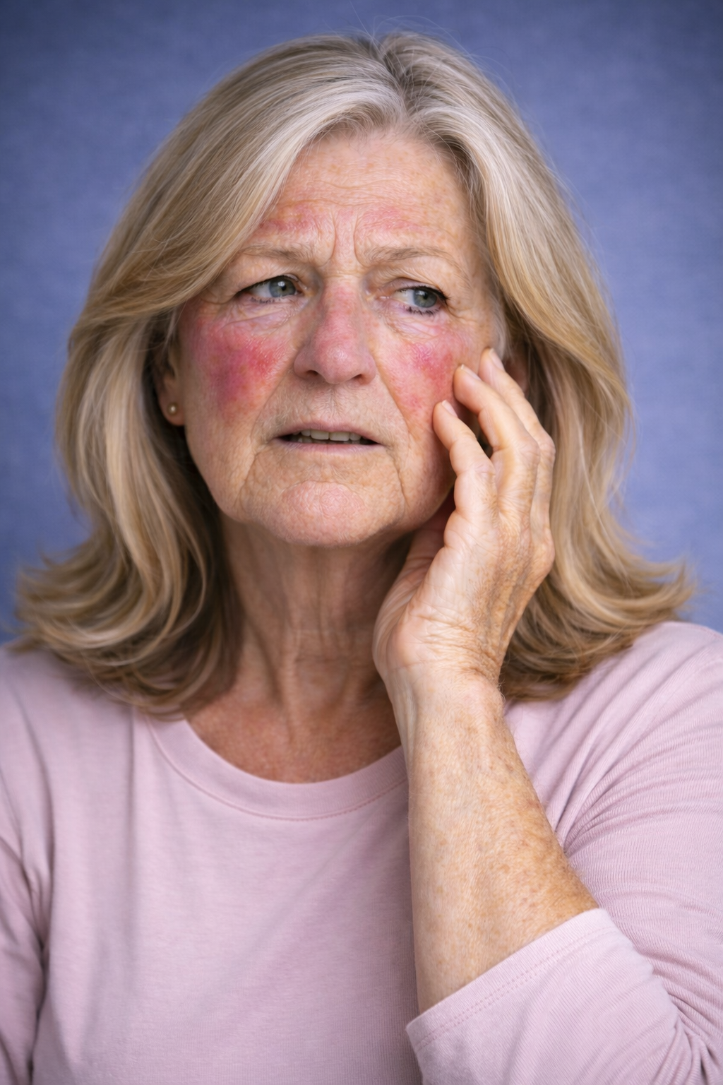
At Village Dermatology in Katy, Texas and Houston, Texas, we often see patients who are concerned about sudden changes in their skin tone. A 40-year-old woman recently came to our office worried about darker patches developing on both cheeks over the past several weeks.
Her main question during the visit was:
“Why are dark patches showing up on my cheeks after my eyelid rash?”
Let’s break down what was happening and how we treated it.
Case Overview: Skin Discoloration on the Cheeks
This patient presented with:
Darker-than-normal skin patches on the right and left cheeks
Moderate discoloration
Symptoms present for several weeks
A recent history of rash around the eyelids
On examination with dermoscopy, we noted ill-defined hyperpigmented patches consistent with:
Post-Inflammatory Hyperpigmentation (PIH)
The discoloration was secondary to inflammation from her recent eyelid dermatitis.
What Is Post-Inflammatory Hyperpigmentation (PIH)?
Post-inflammatory hyperpigmentation occurs when the skin produces excess pigment after inflammation or irritation.
Common triggers include:
When inflammation resolves, it may leave behind:
Dark brown patches
Gray-brown discoloration
Uneven skin tone
PIH is especially common in individuals with medium to darker skin tones but can affect all skin types.
Why Did the Eyelid Rash Cause Cheek Discoloration?
This patient also reported:
Rash under the eyes
Onset during seasonal allergy flare
No new skincare products
She was diagnosed with:
Eyelid Dermatitis
Eyelid skin is extremely thin and sensitive. Allergies, rubbing, and inflammation can trigger dermatitis, which may spread or cause pigment changes in nearby areas like the cheeks.
Even after the rash improves, pigmentation can linger for months.
Treatment Plan at Village Dermatology
At Village Dermatology in Katy and Houston, Texas, we treat both the inflammation and the pigmentation to restore healthy, even skin tone.
1. Treat the Eyelid Dermatitis
We prescribed:
Elidel (pimecrolimus) 1% cream
Apply once daily to eyelids
Elidel is a non-steroidal anti-inflammatory cream that is safe for delicate eyelid skin.
We also recommended:
Daily Zyrtec for allergy control
Discontinuing all skincare products temporarily
Using only hypoallergenic, fragrance-free products
If no improvement, we discussed the possibility of switching to Opzelura.
2. Treat the Post-Inflammatory Hyperpigmentation
For pigmentation management, we recommended:
Broad Spectrum Sunscreen SPF 30+ (daily use)
Sun protective clothing
Topical retinoids to encourage skin cell turnover
Sun exposure can worsen hyperpigmentation and delay fading. Consistent sunscreen use is critical.
How Long Does Post-Inflammatory Hyperpigmentation Last?
We counseled the patient that:
PIH can take months to years to fully fade
Improvement is gradual
Early treatment improves outcomes
Strict sun protection is essential
Patience and consistent treatment are key.
When Should You See a Dermatologist for Facial Discoloration?
You should seek evaluation if:
Dark patches appear suddenly
Pigmentation follows a rash
Discoloration is spreading
Over-the-counter creams are not working
The rash keeps recurring
Proper diagnosis is important because facial pigmentation can also represent:
Melasma
Lichen planus pigmentosus
Drug reactions
Chronic dermatitis
Expert Pigmentation and Eyelid Rash Treatment in Katy and Houston, Texas
At Village Dermatology, we specialize in:
Post-inflammatory hyperpigmentation treatment
Eyelid dermatitis management
Facial discoloration
Allergy-related skin conditions
Medical dermatology for adults
If you are experiencing dark patches, eyelid rash, or uneven skin tone in Katy, Texas or Houston, Texas, our dermatology team is here to help.
Schedule your consultation today and restore clarity and confidence to your skin.
Why Did I Suddenly Break Out in an Itchy Rash on My Face?
A 74-year-old woman presented to Village Dermatology in Katy, Texas with a sudden itchy facial rash diagnosed as allergic contact dermatitis. Learn how expert dermatologic care in Houston and Katy can help treat and prevent facial rashes.
At Village Dermatology in Katy, Texas and Houston, Texas, we frequently see patients who are alarmed by sudden facial rashes. A 74-year-old female recently came to our office as a new patient with a one-week history of a severely itchy facial rash.
Her main concern was simple and urgent:
“Why did I suddenly break out in an itchy rash on my face?”
Here’s what we discovered — and how we helped.
Sudden Facial Rash in a 74-Year-Old Woman
The patient reported:
Sudden onset of itching on the face
Moderate severity
Rash present for one week
Severe pruritus (itching) at its peak
Recent treatment with oral prednisone
Use of over-the-counter Benadryl cream
Using dermoscopic evaluation and reviewing submitted photographs taken during the peak of the eruption, the clinical findings were most consistent with:
Allergic Contact Dermatitis (ACD)
What Is Allergic Contact Dermatitis?
Allergic contact dermatitis is a delayed hypersensitivity reaction that occurs when the skin comes into contact with a substance that triggers an immune response.
Common triggers include:
New skincare products
Fragrances
Cosmetics
Hair dyes
Nail products
Sunscreens
Metals (nickel)
Plants
Scented soaps
Sometimes the reaction appears days after exposure, making it difficult to identify the culprit.
Why Patch Testing Was Delayed
Because the patient was currently taking oral prednisone, we advised that patch testing must be postponed for at least three weeks.
Steroids can suppress immune responses and cause false-negative patch testing results.
In the meantime, we began insurance verification so testing can be scheduled promptly once she is eligible.
Patch testing is often critical when:
The rash persists
The trigger is unclear
Multiple potential allergens are involved
Treatment Plan for Allergic Contact Dermatitis
At Village Dermatology in Katy and Houston, we focus on both immediate relief and long-term prevention.
Immediate Treatment
Apply twice daily for two weeks
Then use as needed for flares
Topical steroids help calm inflammation and reduce itching.
Essential Skincare Reset
We advised the patient to:
Discontinue all current skincare products
Avoid fragrances and scented products
Use only hypoallergenic, unscented soaps
Avoid new cosmetics, shampoos, and sunscreens
Use gentle moisturizers
When allergic contact dermatitis occurs, simplifying your skincare routine is crucial.
Additional Finding: Facial Ecchymosis from CPAP Mask
During examination, we also noted non-palpable purpuric patches (bruising) on both cheeks.
These findings were consistent with:
Ecchymosis (Bruising)
In this case, likely caused by pressure from the patient’s CPAP mask.
Patients using CPAP devices may develop:
Pressure-related bruising
Skin fragility
Friction-related irritation
We recommended:
Loosening or refitting the CPAP mask
Applying a barrier cream such as Desitin to reduce friction
Monitoring for worsening bruising
Most ecchymoses resolve within 3–4 weeks without treatment.
When Should You See a Dermatologist for a Facial Rash?
Seek evaluation if:
A rash appears suddenly without explanation
Itching is severe
Over-the-counter treatments are not helping
The rash spreads or worsens
You suspect a product reaction
Facial skin is delicate, and misdiagnosis can prolong symptoms.
Early dermatologic evaluation helps prevent chronic inflammation and skin damage.
Expert Rash Treatment in Katy and Houston, Texas
At Village Dermatology, we specialize in diagnosing and treating:
Allergic contact dermatitis
Facial rashes
Itchy skin conditions
CPAP-related skin irritation
Chronic inflammatory skin disorders
If you are experiencing a sudden facial rash in Katy, Texas or Houston, Texas, our board-certified dermatology team is here to help.
Schedule your consultation today and let us help restore your skin’s health and comfort.

Why Is My Face So Red, Flaky, and Itchy — and Why Won’t It Go Away?
An 80-year-old male presented to Village Dermatology in Katy, Texas with a chronic red, flaky facial rash diagnosed as seborrheic dermatitis. Learn how expert dermatologic care in Houston and Katy can effectively manage persistent facial rashes.
At Village Dermatology in Katy, Texas and Houston, Texas, we frequently see patients who are frustrated by persistent facial rashes. Recently, an 80-year-old gentleman came to our office with concerns about a red, itchy, flaking rash on his face that had been present for several months.
He asked a question we hear often:
“Why is my face so red, flaky, and itchy — and why won’t it go away?”
Let’s break down what was happening and how we helped.
Understanding Chronic Facial Rashes in Older Adults
This patient reported:
Persistent redness on the forehead
Flaking and scaling skin
Itching and irritation
Symptoms lasting for months
On examination using dermoscopy, we noted pink-to-orange scaly plaques distributed across:
The right inferior forehead
The left inferior forehead
The posterior mid-parietal scalp
Based on the clinical presentation, the diagnosis was seborrheic dermatitis.
What Is Seborrheic Dermatitis?
Seborrheic dermatitis is a chronic inflammatory skin condition that commonly affects:
Face (especially eyebrows, forehead, sides of nose)
Scalp
Ears
Beard area
It presents with:
Redness
Flaking
Greasy or dry scale
Itching
It is extremely common in older adults and can flare due to:
Stress
Illness
Changes in environment
Underlying medical conditions
Many patients in assisted living or transitional care facilities experience flares due to environmental changes and stress on the body.
The important thing to understand is:
Seborrheic dermatitis is chronic. It has periods of flares and remissions. It can be controlled, but not permanently cured.
Treatment Plan for Facial Seborrheic Dermatitis
At Village Dermatology in Katy and Houston, we focus on creating simple, practical treatment regimens that are easy to follow.
For this patient, we recommended:
1. Medicated Shampoo
Ketoconazole 2% shampoo
Use 2–3 times per week during flares
Leave on for 5–10 minutes before rinsing
Use once weekly for maintenance
This helps reduce yeast overgrowth that contributes to inflammation.
2. Combination Topical Therapy for Flares
During flare-ups:
Mix ketoconazole cream with hydrocortisone 2.5% cream
Apply to affected areas on the face and neck twice daily
Use for 2 weeks only during flares
We carefully counsel patients that prolonged steroid use on the face can cause:
Skin thinning
Lightening of the skin
Visible small blood vessels
Using low-potency steroids appropriately and only during flares minimizes these risks.
3. Gentle Skin Care Routine
We advised:
Dove Sensitive Skin body wash
Daily moisturizing with CeraVe or Vanicream
Avoid harsh soaps or scrubs
Gentle skincare is essential for managing seborrheic dermatitis long term.
Additional Concerns Addressed During the Visit
Onychomycosis (Toenail Fungus)
The patient had longstanding toenail fungus. We discussed that treatment is optional if it is not bothersome.
Key counseling points:
Oral medications are more effective but carry potential side effects.
Topical treatments (like OTC Lamisil) may help if fungus spreads to surrounding skin.
Lower Extremity Ulcers
The patient also had ulcers on the right great toe and left ankle following surgery. Because he has diabetes and neuropathy, we emphasized:
Close wound care follow-up
Monitoring for infection
Continued coordination with wound care specialists
Fortunately, there were no signs of infection during the visit.
When Should You See a Dermatologist for a Facial Rash?
You should schedule an evaluation if:
The rash lasts more than a few weeks
Over-the-counter products are not helping
The rash spreads
There is significant itching or discomfort
You are unsure of the diagnosis
Many patients assume persistent facial redness is just “dry skin.” In reality, it may be seborrheic dermatitis, rosacea, psoriasis, or another inflammatory condition.
An accurate diagnosis makes all the difference.
Expert Seborrheic Dermatitis Care in Katy and Houston, Texas
At Village Dermatology, we specialize in diagnosing and managing chronic skin conditions in patients of all ages — including elderly patients in transitional care and assisted living facilities.
If you or a loved one is experiencing:
Red, flaky facial skin
Chronic scalp scaling
Persistent itching
Recurrent facial rashes
We are here to help.
Schedule an appointment at Village Dermatology in Katy, Texas or Houston, Texas today.
Clearer, healthier skin is possible — even with chronic conditions.
Melasma Treatment Follow-Up in Katy & Houston, TX: Case Report of Persistent Facial Hyperpigmentation
A 50-year-old male followed up for melasma treatment with tretinoin and oral tranexamic acid. Learn how Village Dermatology in Katy and Houston, Texas manages persistent facial hyperpigmentation.
By: Dr. Caroline Vaughn
Melasma can be one of the most frustrating pigment conditions to treat due to its chronic, relapsing nature. At Village Dermatology, we take a multimodal and maintenance-based approach to help patients achieve long-term improvement. This case highlights a melasma follow-up visit in a 50-year-old male treated in Katy and Houston, Texas.
Patient Presentation
A 50-year-old male returned for a three-month follow-up for melasma affecting the left central malar cheek.
At his previous visit (August 27, 2025), he was prescribed:
Tretinoin 0.025% cream (pea-sized amount, 2–3 nights weekly increasing to nightly as tolerated)
Oral tranexamic acid 650 mg, ½ tablet twice daily
Despite adherence to treatment, the patient reported minimal improvement. He noted that a prior compounded hydroquinone formulation had provided slightly better results.
Clinical Examination
A focused facial examination was performed using dermoscopy.
Findings included:
Ill-defined hyperpigmented patches
Periorbital/malar distribution
Located primarily on the left central malar cheek
Overall, the melasma appeared relatively controlled but with persistent residual pigmentation.
Diagnosis
Melasma
Chronic hyperpigmentation disorder affecting sun-exposed facial areas.
Treatment Plan
After extensive discussion, the following plan was agreed upon:
Restart Compounded Hydroquinone
Resume a 3-month course
Emphasized that hydroquinone is not intended for long-term continuous use
Reviewed risks including rare pseudoochronosis
Continue Tretinoin 0.025%
Safe for long-term maintenance therapy
Apply pea-sized amount nightly as tolerated
Procedural Options Discussed
Melanage® peel
Superficial chemical peels
Other pigment-targeting treatments
The patient may consider a Melanage peel in the future.
Importance of Sun Protection
Strict daily photoprotection was emphasized as the foundation of melasma management:
Reapplication during sun exposure
Sun avoidance when possible
Protective hats and clothing
Without consistent sun protection, melasma recurrence is common.
Patient Counseling & Education
Expectations
Melasma is chronic and often triggered by:
Sun exposure
Hormonal changes
Heat
Maintenance therapy is typically required
Retinoid Counseling
Apply at bedtime
Wait 30 minutes after washing face
Use moisturizer if dryness occurs
When to Contact the Office
If melasma worsens
If medication side effects develop
If pigment changes appear concerning
Clinical photos were obtained for monitoring progress. The patient will message the office if he needs a refill on compounded hydroquinone.
Follow-Up
Follow up in 4 months for reassessment
Long-term maintenance visit annually
Advanced Melasma Treatment in Katy & Houston
At Village Dermatology, we provide comprehensive melasma management including:
Topical retinoids
Hydroquinone therapy
Oral tranexamic acid
Chemical peels
Laser treatments
If you are struggling with facial hyperpigmentation or dark patches, our dermatology team in Katy and Houston, Texas can help create a customized treatment plan.
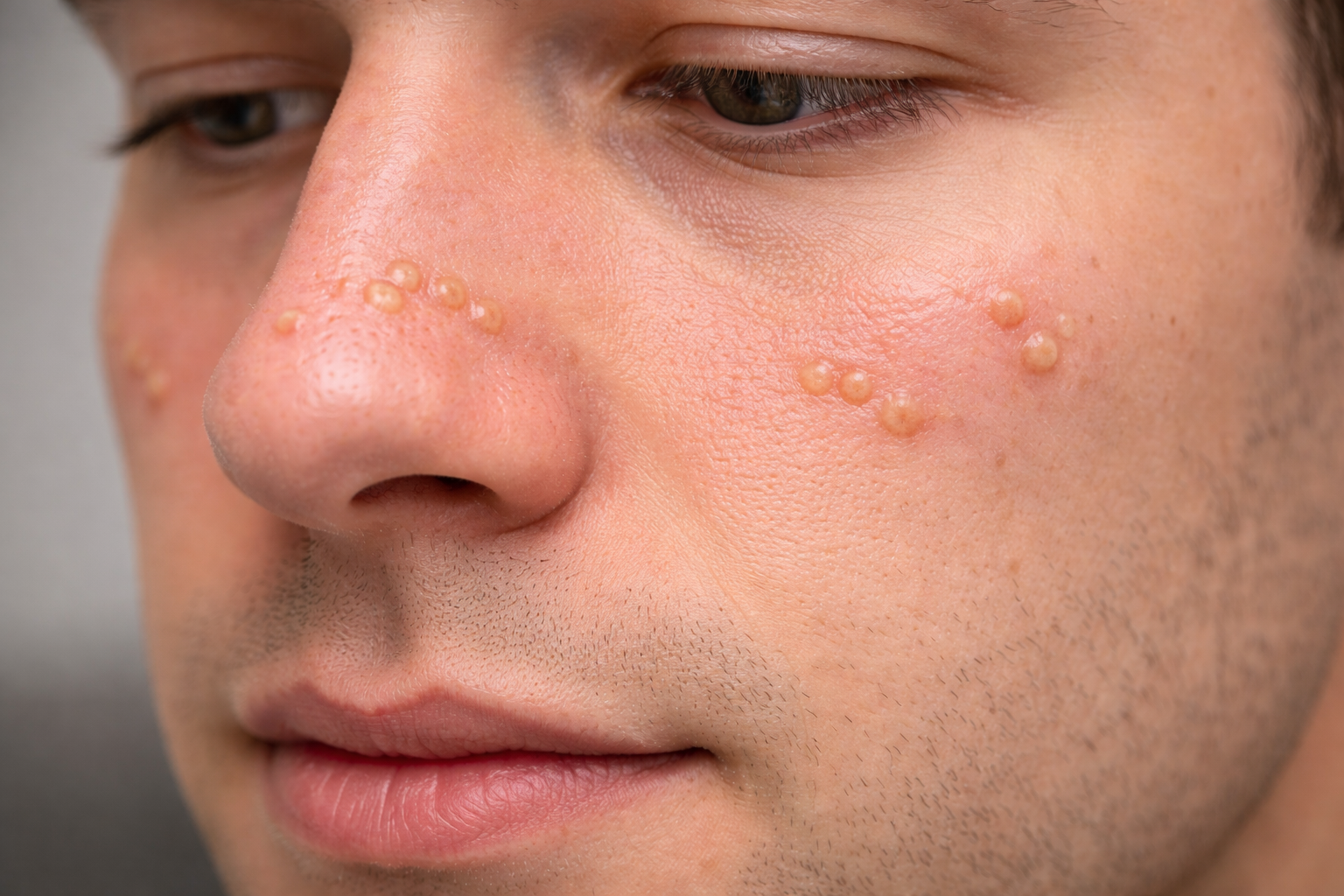
Sebaceous Hyperplasia Treatment Case Report: Cosmetic Lesion Removal in Katy & Houston, TX
A 22-year-old male underwent cosmetic electrodesiccation for sebaceous hyperplasia on the nose and cheeks. Learn how Village Dermatology in Katy and Houston, Texas treats benign facial lesions safely and effectively.
By: Dr. Ashley Baldree
Facial skin lesions that darken or enlarge can understandably cause concern, especially in young adults. At Village Dermatology, we carefully evaluate all changing facial lesions to rule out concerning pathology and provide safe, effective cosmetic treatment options. This case highlights the diagnosis and cosmetic removal of sebaceous hyperplasia in a 22-year-old patient in Katy and Houston, Texas.
Patient Presentation
A 22-year-old male, an established patient, presented for evaluation of multiple facial skin lesions located on:
Nose
Right cheek
Left cheek
The lesions had been present for several months and were described as:
Darkening
Enlarging
Irregular in appearance
Moderate in severity
The lesions had not been previously treated. The patient requested evaluation and management.
Clinical Examination
A focused dermatologic examination of the scalp, head, and face was performed.
Findings revealed multiple small papules distributed on:
Nasal dorsum
Right nasal sidewall
Nasal tip and supratip
Left superior medial malar cheek
Left medial malar cheek
The appearance was consistent with sebaceous hyperplasia, a benign enlargement of oil glands.
Diagnosis
Sebaceous Hyperplasia
Benign enlarged sebaceous (oil) glands of the face.
Treatment Plan
The benign nature of sebaceous hyperplasia was reviewed with the patient. Treatment options were discussed, including:
Electrodessication
Laser therapy
The patient elected to proceed with cosmetic electrodesiccation.
Procedure Details
10 lesions treated
Locations: nasal dorsum, bilateral malar cheeks, nasal sidewall, tip, and supratip
Informed consent obtained
Risks reviewed, including:
Crusting
Scabbing
Blistering
Pigment changes
Recurrence
Infection
Scarring
Detailed post-procedure care instructions were provided.
Post-Procedure Instructions
Apply Vaseline to treated areas if crusting occurs
Avoid picking or scratching
Use daily sun protection (SPF 30+)
Expect mild redness and scabbing during healing
Patients were instructed to contact the office if lesions fail to improve or if signs of infection develop.
Additional Cosmetic Consultation
The patient also consulted regarding acne scarring, with plans to consider treatment after completion of his isotretinoin (Accutane®) course.
Potential future treatments discussed included:
CO₂ laser resurfacing
Subcision
Chemical peels
Cosmetic Dermatology in Katy & Houston
Sebaceous hyperplasia is common in young adults and can mimic more concerning lesions. At Village Dermatology, we provide expert facial lesion evaluation and advanced cosmetic treatments, including electrodesiccation and laser therapy, in Katy and Houston, Texas.
If you notice enlarging or changing facial lesions, schedule a professional evaluation for peace of mind.
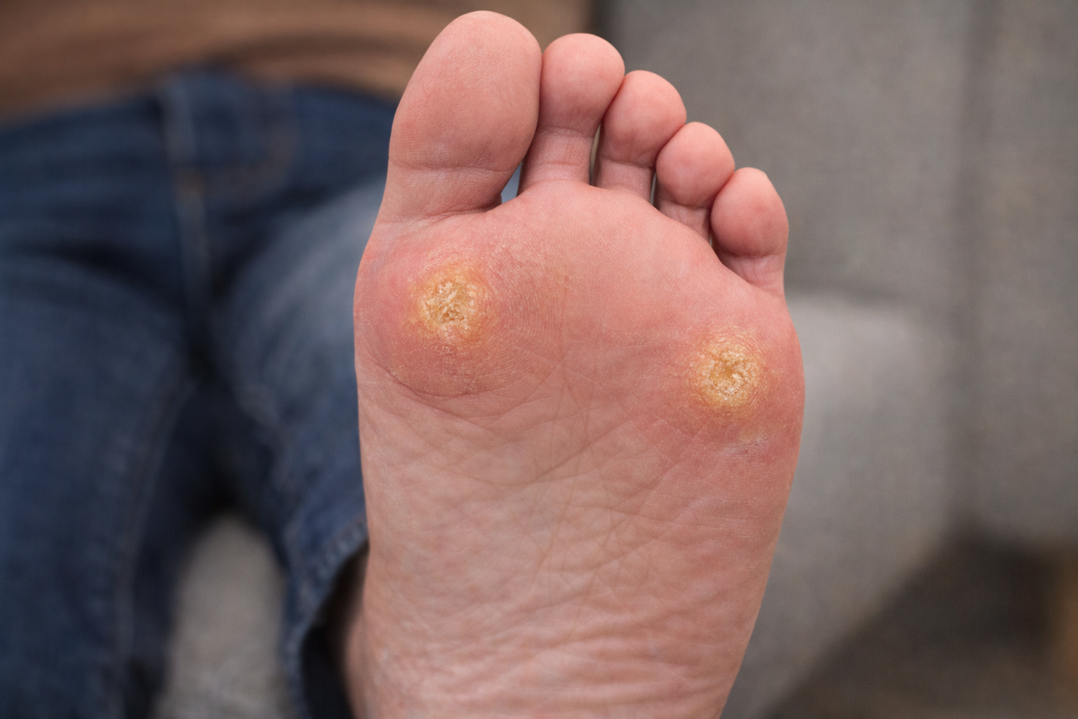
Plantar Callus Treatment Case Report: Managing Hyperkeratotic Foot Lesions in Katy & Houston, TX
A 38-year-old male was evaluated for plantar calluses on the foot. Learn how Village Dermatology in Katy and Houston, Texas treats hyperkeratotic foot lesions using urea 40% and keratolytic therapy.
By: Dr. Ashely Baldree
Thickened skin on the soles of the feet is a common concern, especially in active adults. At Village Dermatology, we frequently evaluate lesions on the plantar surface to determine whether they represent benign calluses, warts, or more concerning growths. This case highlights the evaluation and treatment of plantar calluses in a patient seen in Katy and Houston, Texas.
Patient Presentation
A 38-year-old male presented as a new patient for evaluation of a skin lesion on the right plantar surface of the foot. The lesion had been present for several months and was:
Asymptomatic
Stable in size
Not previously treated
Not associated with a family history of melanoma
The patient requested evaluation to ensure the lesion was benign and to discuss treatment options.
Clinical Examination
A focused dermatologic examination of both feet was performed using dermoscopy.
Findings included:
Hyperkeratotic plaques
Located on pressure-bearing areas:
Right medial plantar midfoot
Left medial plantar heel
The appearance and distribution were consistent with plantar calluses caused by friction and pressure rather than a neoplastic process.
Assessment
Callus
Hyperkeratotic plaques on pressure-bearing surfaces of the feet.
Treatment Plan
The patient was reassured that the lesions were benign calluses resulting from chronic friction.
Recommended Treatment:
Urea 40% topical cream, applied nightly to the soles
Samples of urea lotion provided in-office
Continue use of keratolytic agents such as:
Amlactin®
Duofilm®
Mediplast®
Urea 40% helps soften and break down thickened skin, improving texture and comfort over time.
Patient Counseling & Education
Why Calluses Form
Calluses develop due to:
Repeated friction
Pressure from footwear
Abnormal weight distribution
Expectations
Improvement typically occurs with consistent keratolytic therapy
Reduction of friction (proper footwear, insoles) is key to prevention
Calluses may recur if pressure persists
When to Contact the Office
If lesions worsen
If pain develops
If no improvement after several months of treatment
Follow-Up
Follow up as needed if symptoms worsen or fail to improve.
Expert Foot & Skin Care in Katy & Houston
At Village Dermatology, we evaluate all plantar lesions carefully to rule out warts, atypical growths, or precancerous changes. Whether you're dealing with calluses, dry cracked heels, plantar warts, or other foot skin conditions, our dermatology team provides personalized, evidence-based treatment.
If you have thickened or persistent skin lesions on your feet, schedule an evaluation with Village Dermatology in Katy or Houston, Texas.
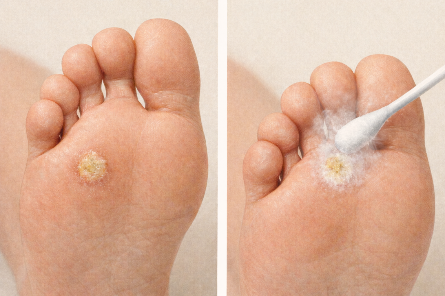
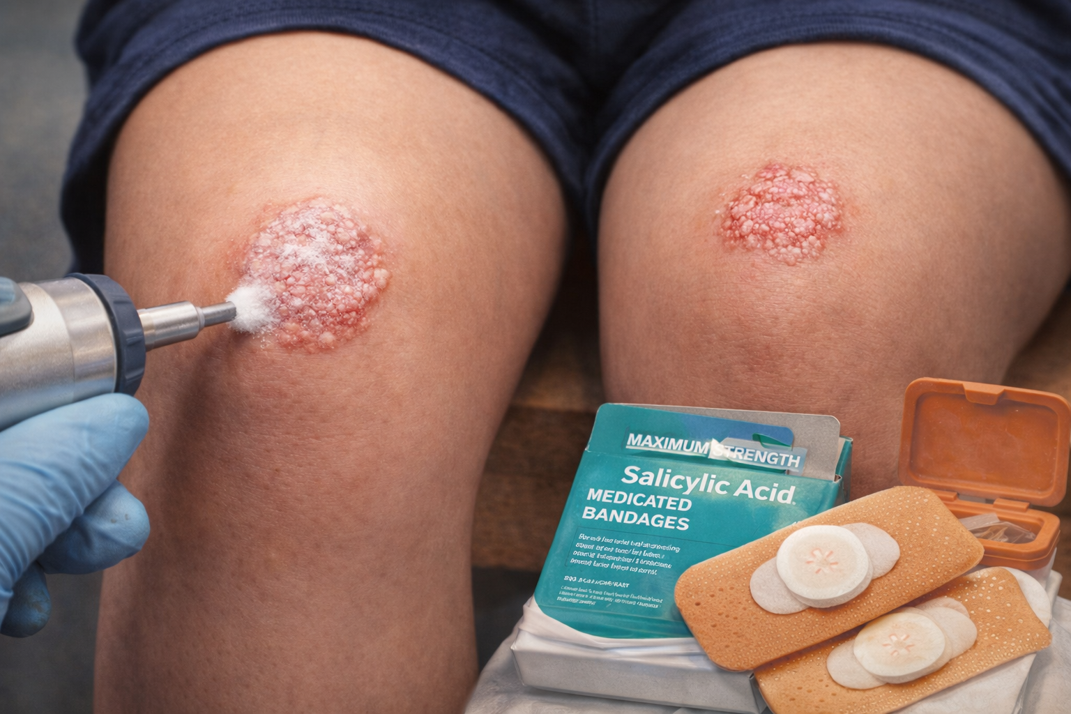
Treating a Painful Plantar Wart: A Case Study from Village Dermatology in Katy & Houston, Texas
A 38-year-old male with a painful plantar wart on the left foot underwent paring and liquid nitrogen treatment at Village Dermatology in Katy & Houston, Texas. Learn about plantar wart causes, treatments, and recovery expectations.
Plantar warts are a common but often stubborn skin condition that can significantly impact daily activities—especially for active individuals. At Village Dermatology in Katy and Houston, Texas, we frequently treat plantar warts that have not responded to over-the-counter therapies.
This case highlights a 38-year-old male who presented with a persistent and painful plantar wart on the left foot, requiring in-office procedural treatment for relief.
Patient Overview
Chief Complaint
Wart on the left plantar surface of the foot
Present for approximately 2–3 months
Moderate severity
Causing discomfort during workouts
Previous Treatments
Over-the-counter salicylic acid
OTC cryotherapy
Partial improvement only; lesion persisted
Clinical Examination
A focused foot exam revealed:
A plantar wart on the left lateral plantar midfoot
Hyperkeratotic lesion consistent with verruca plantaris
No signs of secondary infection
Patient otherwise healthy, well-nourished, and in no distress
Diagnosis: Plantar Wart (Verruca Plantaris)
Plantar warts are caused by the human papillomavirus (HPV) and occur on the soles of the feet. Due to the thick skin in this area and constant pressure from walking, plantar warts are among the most treatment-resistant warts.
They can:
Be painful
Spread with direct contact
Persist for months to years without proper treatment
Treatment Plan
In-Office Procedure: Paring + Liquid Nitrogen (Cryotherapy)
During the visit:
The lesion was pared with a curette to remove thickened skin
Liquid nitrogen (LN2) was applied to the wart
One lesion treated during this session
Patient Consent & Education
The patient was counseled and consented regarding potential risks, including:
Blistering
Crusting or scabbing
Pigment changes
Scarring
Recurrence or incomplete removal
Infection
The patient tolerated the procedure well.
Counseling & Expectations
The patient was advised:
Plantar warts often require 3–4 liquid nitrogen treatments for full resolution
Treatments are typically spaced every 3–4 weeks
Discomfort after treatment is common but temporary
At-Home Care
Continue topical salicylic acid between visits
Avoid picking or shaving the lesion
Keep feet clean and dry
Wear protective footwear in communal areas (gyms, locker rooms, pools)
When to Contact the Office
If the wart spreads
If it recurs after treatment
If pain or signs of infection develop
Follow-Up Plan
Return in 1 month for re-evaluation and possible repeat cryotherapy
Expert Wart Treatment in Katy & Houston, Texas
Plantar warts can be frustrating, painful, and difficult to treat without professional care. At Village Dermatology, we offer a wide range of evidence-based treatments including:
Cryotherapy (liquid nitrogen)
Cantharidin
Salicylic acid therapy
Candidal antigen injections
Laser therapy
Surgical options when necessary
Our dermatology team customizes treatment based on lesion location, size, symptoms, and patient lifestyle.
Comprehensive Skin Evaluation and Preventive Counseling
Case report from Village Dermatology in Katy and Houston, TX highlighting evaluation and punch biopsy of a darkly pigmented ear lesion, benign skin findings, and comprehensive skin cancer prevention counseling.
Village Dermatology | Katy & Houston, Texas
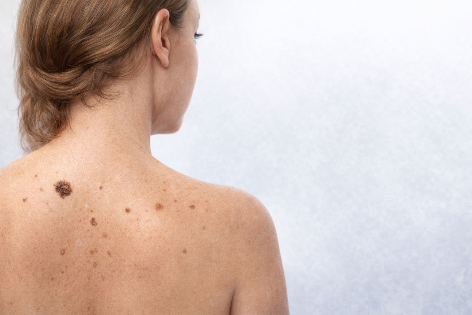
In addition to evaluating the concerning pigmented lesion on the ear, the patient underwent a comprehensive full-body skin examination (FBSE) at Village Dermatology. Multiple benign skin findings were identified, and detailed counseling was provided to support long-term skin health and cancer prevention.
Benign Skin Findings and Counseling
Seborrheic Keratoses
Seborrheic keratoses were identified and discussed with the patient.
Patient Education:
Seborrheic keratoses are benign, non-cancerous growths
They often appear as warty or “stuck-on” lesions
These growths commonly increase with age
Plan:
Reassurance and counseling only. No treatment was required.
Cherry Angiomas
Diagnosis: Cherry angiomas
Location: Right superior medial upper back
Patient Education:
Cherry angiomas are benign vascular growths
Treatment is not medically necessary
Cosmetic treatment options include laser therapy or electrodesiccation if desired
Plan:
Counseling and reassurance.
Lentigines
Diagnosis: Lentigines
Location: Left superior medial upper back
Patient Education:
Lentigines are benign pigmented lesions commonly related to sun exposure
They frequently occur on sun-damaged skin
These lesions are highly treatable
Treatment Options Discussed:
Broad-spectrum sunscreen
Sun avoidance
Bleaching creams
Retinoids
Chemical peels
Laser treatments
Plan:
Counseling with emphasis on sun protection.
Sun Protection Counseling
Given the patient’s sun-related skin findings and family history, comprehensive sunscreen education was provided.
Recommendations:
SPF 30 blocks approximately 97% of harmful UV rays
Apply sunscreen 15 minutes before sun exposure
Reapply every 2 hours, or every 45–60 minutes when swimming or sweating
Use approximately one ounce (shot glass amount) to cover exposed skin
Use lip balm with SPF
Sun-protective clothing is an effective alternative when worn consistently
Acrochordons (Skin Tags)
Diagnosis: Acrochordons
Location: Right inferior anterior neck
Patient Education:
Skin tags are benign skin growths
Commonly occur on the neck and underarms
Can become irritated by clothing or jewelry
Treatment Options:
Surgical removal
Liquid nitrogen if symptomatic or cosmetically bothersome
Plan:
Counseling and reassurance.
Family History of Malignant Melanoma
Risk Factor: Father deceased from malignant melanoma (Z80.8)
Patient Counseling:
A first-degree relative with melanoma increases personal risk
Monthly self-skin examinations are essential
Watch for moles that change in size, shape, or color, or that itch, bleed, or burn
Daily sun protection and protective clothing are critical
Instructions:
Contact the office immediately for any new or changing lesions.
Preventive Health & Quality Measures (MIPS)
The following quality measures were addressed:
Tobacco Use Screening: Patient is an ex/non-smoker
Alcohol Use Screening: No unhealthy alcohol use identified
Medication Reconciliation: Current medications documented
Follow-Up Plan
The patient was advised to return in one year for a full-body skin examination (FBSE) or sooner if any new or changing skin lesions are noted.
Why This Matters
This case underscores the importance of early evaluation, biopsy when indicated, routine skin checks, and patient education—especially for individuals with a family history of melanoma. Early diagnosis truly saves lives.
At Village Dermatology, we are proud to provide expert, compassionate dermatologic care to patients in Katy, Houston, and surrounding Texas communities.
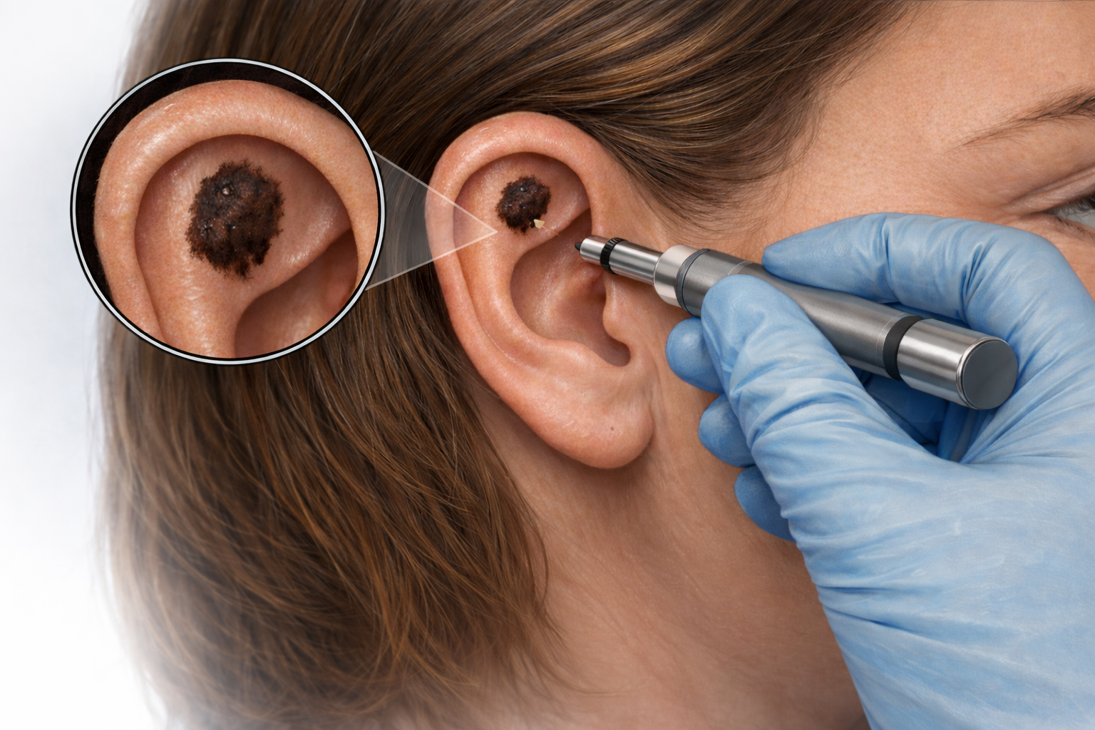
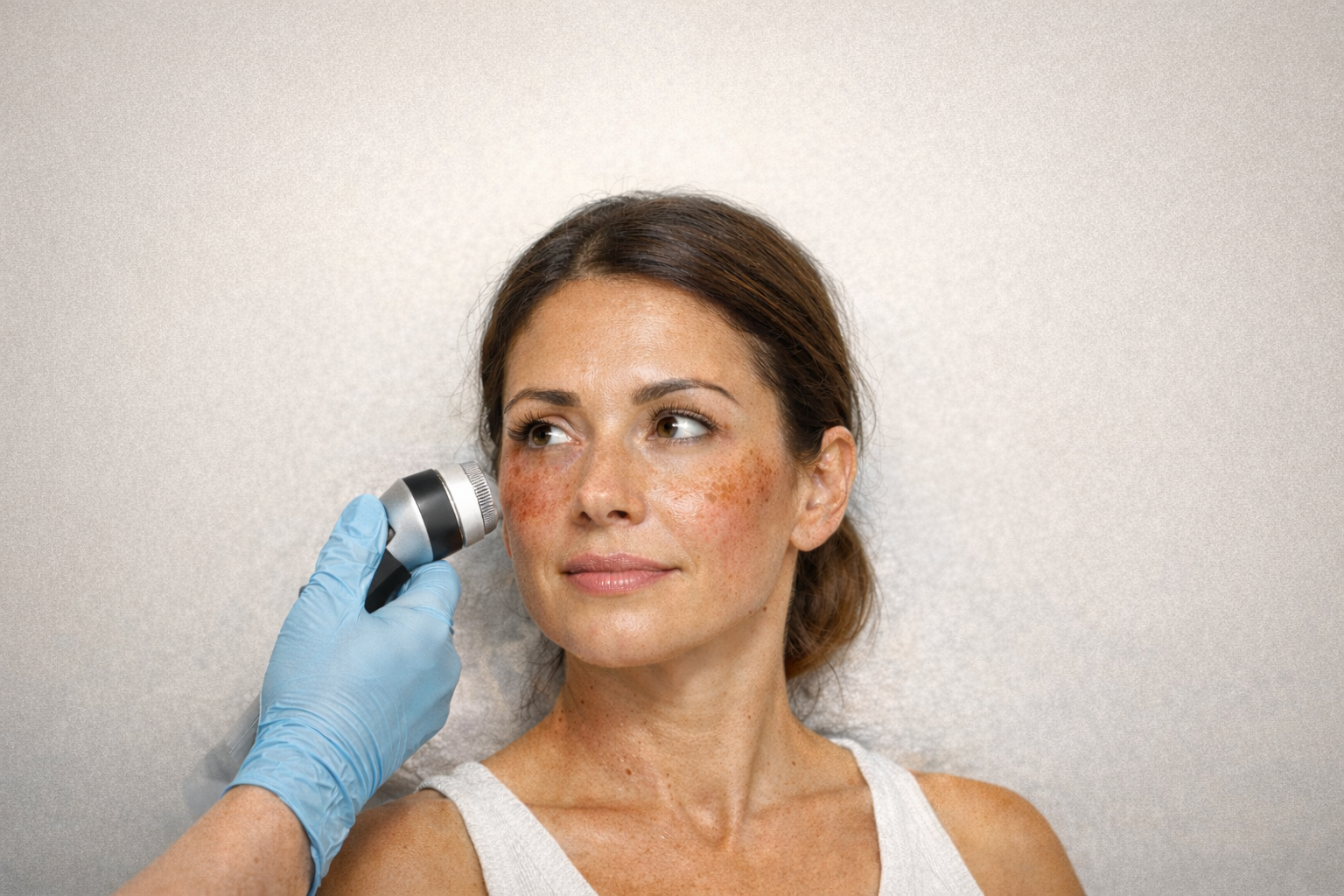
Darkening Lesion on the Ear: Why Early Evaluation Matters
A changing, darkly pigmented lesion on the ear was evaluated and biopsied at Village Dermatology in Katy and Houston, Texas. Learn why early skin checks matter.
Village Dermatology | Katy & Houston, Texas
A changing skin lesion should never be ignored—especially when it appears on sun-exposed areas like the ears. At Village Dermatology, we frequently evaluate concerning skin lesions to ensure early diagnosis and appropriate management.
In today’s case report, we highlight the evaluation and biopsy of a darkly pigmented lesion on the ear in an established patient.
Patient Presentation
A 58-year-old female presented to our dermatology clinic with concerns about a skin lesion on the right ear. She reported that the lesion had been present for several months and had gradually become darker, larger, and more irregular in appearance. The lesion had not been treated previously.
Because of the lesion’s location and changes over time, the patient was seen for prompt evaluation and management.
Dermatologic Examination
A focused skin examination was performed, including evaluation of the scalp, face, and upper extremities. The patient appeared well-developed, well-nourished, and in no acute distress.
Using dermatoscopy, a darkly pigmented macule was identified on the right antihelix of the ear. Dermatoscopic examination allows dermatologists to better assess pigment patterns and structural features that are not visible to the naked eye.
Clinical Impression and Differential Diagnosis
Based on the lesion’s appearance and evolution, the clinical impression was:
Neoplasm of Unspecified Behavior
The differential diagnosis included:
Neoplasm of unspecified behavior
Chondrodermatitis nodularis helicis (CNH)
Cyst
Given the uncertainty and concerning features, a biopsy was recommended to obtain a definitive diagnosis.
Procedure: Punch Biopsy of the Ear
After discussing risks and benefits, written informed consent was obtained. The biopsy was performed as follows:
Location: Right antihelix
Anesthesia: 1% lidocaine with epinephrine
Technique: 4 mm punch biopsy
Specimen: Sent for histopathologic evaluation (H&E staining)
Closure: 5-0 fast-absorbing gut suture
The patient tolerated the procedure well. Petrolatum and a bandage were applied, and detailed post-procedure care instructions were provided.
Follow-Up and Importance of Biopsy
The patient was advised that she would be notified of the biopsy results and instructed to contact the office if results were not received within two weeks.
This case highlights the importance of early dermatologic evaluation for lesions that are changing in color, size, or shape—particularly in sun-exposed areas like the ears. A simple in-office biopsy can provide critical information and peace of mind.
When to See a Dermatologist
You should schedule a dermatology appointment if you notice:
A mole or lesion that is darkening or enlarging
Irregular borders or uneven color
Lesions on sun-exposed areas such as the ears, face, or scalp
Any skin spot that looks or feels “different”
At Village Dermatology, we proudly serve patients in Katy, Houston, and surrounding Texas communities, offering expert skin cancer screening and personalized dermatologic care.
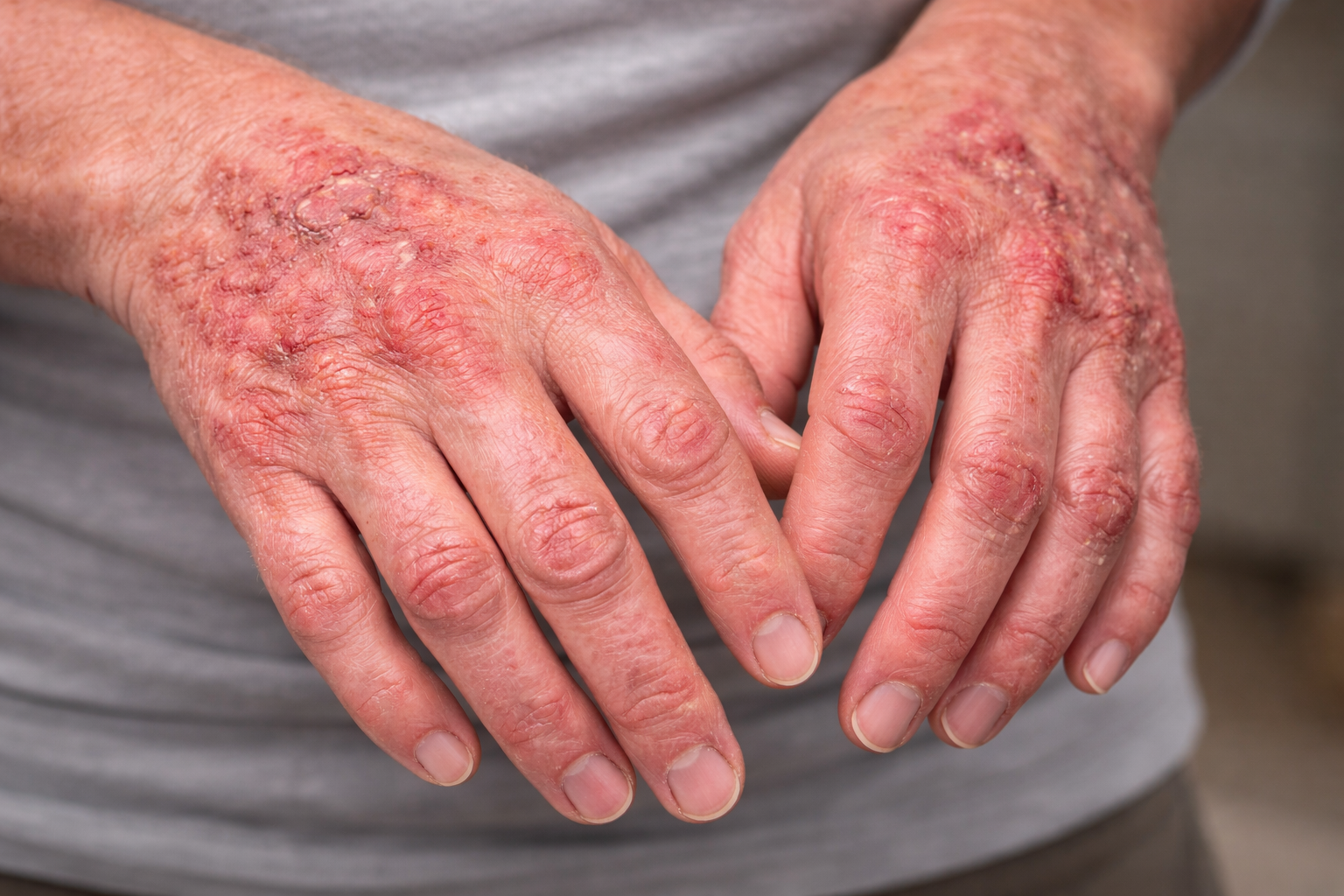
Chronic Hand Dermatitis Case Report: Managing Severe Itching and Fissuring in a 55-Year-Old Female
A 55-year-old female with chronic hand dermatitis and severe itching was treated with high-potency topical steroids and wet wrap therapy. Learn how Village Dermatology in Katy and Houston, Texas manages persistent eczema.
Chronic hand dermatitis can significantly impact daily activities, sleep, and quality of life—especially when symptoms persist despite initial treatment. At Village Dermatology, we specialize in identifying triggers and optimizing treatment plans for inflammatory skin conditions. This case highlights the management of inadequately controlled hand dermatitis in a patient seen in Katy and Houston, Texas.
Patient Presentation
A 55-year-old female presented as a new patient with a 4-month history of a blistering, red, and intensely itchy rash affecting both hands. She reported prior evaluation by her primary care provider and treatment with triamcinolone, which did not provide sufficient relief.
The patient described severe itching, rated 10/10 on the itch numeric rating scale, and noted that the condition was contributing to increased anxiety and sleep disruption.
Clinical Examination
A focused dermatologic examination of the right and left hands was performed using dermoscopy. The patient appeared well-developed, well-nourished, alert, and in no acute distress.
On examination, there were erythematous eczematous patches with fissuring distributed across both hands, consistent with chronic hand dermatitis.
Assessment
Status: Inadequately controlled
Overall severity: Mild with severe pruritus
Treatment Plan
Given the persistence of symptoms and lack of response to mid-potency topical steroids, a more aggressive treatment plan was initiated:
Clobetasol 0.05% ointment, applied twice daily to affected areas on the hands (and feet if involved) for 2–3 weeks
Wet wrap therapy with occlusion using white cotton gloves at night to enhance medication penetration
Hydroxyzine 10 mg orally at bedtime to help relieve itching and improve sleep
Continued use of thick emollient moisturizers multiple times daily
Patient Counseling & Education
Extensive counseling was provided to address both symptom control and long-term management:
Skin Care Recommendations
Wash hands with lukewarm water and a mild, fragrance-free cleanser
Moisturize immediately after washing
Apply emollients 2–3 times daily
Avoid scented soaps, detergents, and fabric softeners
Keep fingernails short
Avoid excessive hand washing when possible
Expectations
The patient was counseled that hand dermatitis is often chronic and relapsing, and may worsen with:
Stress
Dry weather
Frequent hand washing
Harsh or scented products
Skin infections
Medication Counseling
Hydroxyzine may cause drowsiness; patient advised not to drive after taking it
Potential side effects reviewed, including dry mouth, blurry vision, and urinary retention
Risks of prolonged topical steroid use discussed, including skin thinning, discoloration, and visible blood vessels
Patient advised to avoid high-potency steroids on the face, groin, or skin folds
All questions were answered, and the patient demonstrated understanding of the treatment plan.
Follow-Up
Return visit scheduled in 2–3 weeks to assess response to treatment and adjust therapy if needed
Expert Hand Dermatitis Care in Katy & Houston
This case demonstrates the importance of escalation of therapy and patient education when managing chronic hand dermatitis. At Village Dermatology, we provide personalized treatment plans to help patients regain skin comfort and improve quality of life.
If you’re struggling with persistent hand rashes or severe itching, our dermatology team is here to help.
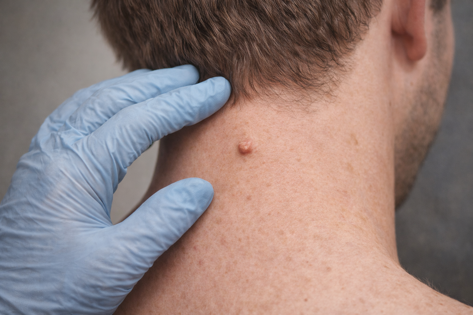
Mole Check & Shave Biopsy Case Report: Evaluating a New Neck Lesion in a 31-Year-Old Male
A 31-year-old male underwent a mole check and shave biopsy for a new neck lesion. Learn how Village Dermatology in Katy and Houston, Texas evaluates and manages suspicious skin growths.
Routine skin checks play a critical role in identifying new or changing lesions early. At Village Dermatology, we emphasize thorough skin evaluations and patient education to ensure timely diagnosis and peace of mind. This case highlights the evaluation and management of a new growth discovered during a routine mole check in Katy and Houston, Texas.
Patient Presentation
A 31-year-old male presented as a new patient for a mole check after his barber noticed a new growth on the back of his neck. The patient denied any personal or family history of melanoma or non-melanoma skin cancer and had no prior history of skin cancer.
He requested evaluation to determine whether the lesion was benign or required further treatment.
Clinical Examination
A focused examination was performed of the scalp, face, head, and neck, with dermoscopy used to further evaluate the lesion. The patient appeared well-developed, well-nourished, alert, and in no acute distress.
On exam, a papule on the left inferior posterior neck was identified. Based on its appearance, the lesion was considered indeterminate.
Assessment
Neoplasm of Uncertain Behavior
Location: left inferior posterior neck
Differential diagnosis included:
Nevus
Acrochordon (skin tag)
Treatment Plan: Shave Biopsy
The risks, benefits, and alternatives were discussed, and the patient elected to proceed with a shave removal biopsy for definitive diagnosis.
Procedure Details
Written consent obtained
Area prepped with alcohol
Local anesthesia achieved using 0.3 cc of 1% lidocaine with epinephrine
Shave biopsy performed to the level of the dermis using a Dermablade
Specimen sent for histopathologic evaluation (H&E)
Hemostasis achieved with Drysol
Petrolatum and bandage applied
The patient was instructed on wound care and advised to contact the office if biopsy results were not communicated within two weeks.
Additional Findings: Skin Tags
Multiple skin tags (acrochordons) were also noted around the neck. These were discussed as benign growths commonly found in friction areas.
Quoted removal of 10 lesions for $150
Counseling provided regarding treatment options, including surgical removal or cryotherapy
Patient Counseling & Education
The patient was counseled on:
Skin cancer awareness and monitoring for new or changing lesions
The benign nature of most nevi and skin tags
When to seek evaluation for concerning changes such as rapid growth, bleeding, or color change
Preventive health screenings were also completed, including tobacco and alcohol use screening.
Follow-Up
Follow up as needed (PRN)
Await pathology results from the shave biopsy
Comprehensive Mole Checks in Katy & Houston
This case underscores the importance of professional skin exams—even for young adults without a personal or family history of skin cancer. At Village Dermatology, we offer thorough mole checks, in-office biopsies, and personalized counseling to help patients stay proactive about their skin health.
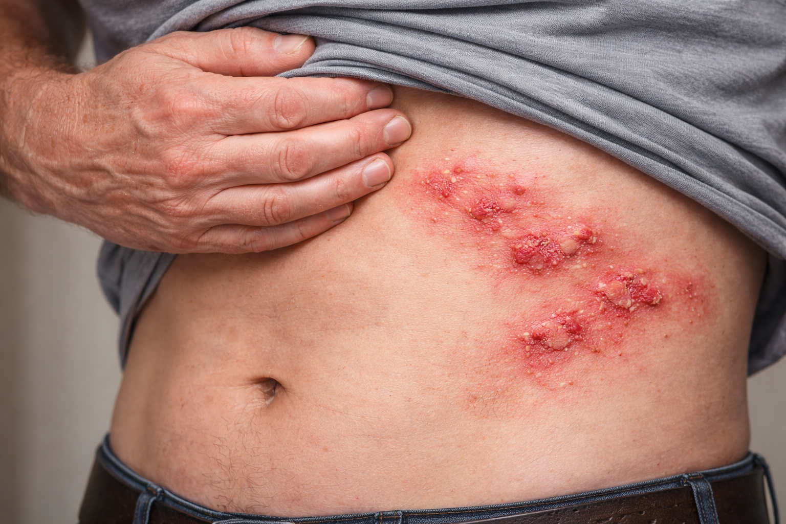
Persistent Rash Case Report: Evaluating Dermatitis and Folliculitis in a 45-Year-Old Male
A 45-year-old male with a persistent rash on the abdomen and hands was evaluated for dermatitis versus folliculitis. Learn how Village Dermatology in Katy and Houston, Texas approaches diagnosis and treatment of complex rashes.
Rashes can be challenging to diagnose when symptoms overlap between inflammatory and infectious skin conditions. At Village Dermatology, we take a comprehensive, stepwise approach to evaluate persistent rashes and tailor treatment plans for optimal outcomes. This case highlights the importance of reassessment, diagnostic testing, and targeted therapy for unresolved skin lesions in Katy and Houston, Texas.
Patient Presentation
A 45-year-old male, an established patient, presented for evaluation of two separate rashes:
Hands: Flaking, itchy rash of moderate severity. The patient had been using Protopic® (tacrolimus).
Trunk (right lateral abdomen): Red, painful lesions associated with burning sensation and intermittent drainage, present since late December 2025. He had completed a 5-day course of Augmentin® and mupirocin ointment, noting partial improvement but persistent lesions.
The patient returned for further evaluation due to incomplete resolution.
Clinical Examination
A focused examination was performed, including the right and left lower extremities. The patient appeared well-developed, well-nourished, alert, and in no acute distress.
On examination, lesions on the right lateral abdomen were consistent with inflammatory and possibly infectious changes, raising concern for:
Dermatitis, unspecified
Folliculitis
Healing ruptured abscess
Assessment
Lesions on the right lateral abdomen
Differential diagnosis: dermatitis vs. folliculitis vs. healing ruptured abscess
Diagnostic Evaluation
Given the persistence of symptoms and drainage, a wound culture was obtained to help guide further management and rule out ongoing infection.
Treatment Plan
To address both inflammatory and potential infectious components, the following treatment plan was initiated:
Doxycycline 100 mg orally twice daily for 10 days
Clindamycin 1% topical gel, applied to affected areas twice daily until improvement
Recommend benzoyl peroxide (BPO) wash or continuation of chlorhexidine wash daily to affected areas
Continue use of emollients and gentle skin care products
Patient Counseling & Education
Extensive counseling was provided, including:
Skin Care
Use gentle cleansers and moisturizers regularly
Avoid harsh or fragranced products
Expectations
The patient was informed that a definitive diagnosis is not always immediate
Empiric therapy and follow-up are sometimes necessary to fully resolve complex rashes
Medication Counseling
Risks of prolonged topical steroid use, including skin thinning, pigment changes, and visible blood vessels
Importance of avoiding high-potency steroids on the face, groin, and skin folds
When to Contact the Office
Development of fever
Rapid worsening of the rash
Increased pain or drainage
All questions were addressed, and the patient demonstrated understanding of the treatment plan.
Follow-Up
Return visit scheduled in 2 weeks for reassessment and review of culture results
Expert Rash & Dermatitis Care in Katy & Houston
This case illustrates the importance of reassessment and diagnostic evaluation when rashes persist despite initial treatment. At Village Dermatology, we provide comprehensive care for complex skin conditions using evidence-based therapies and personalized treatment plans.
If you’re dealing with a persistent or painful rash, our dermatology team is here to help.
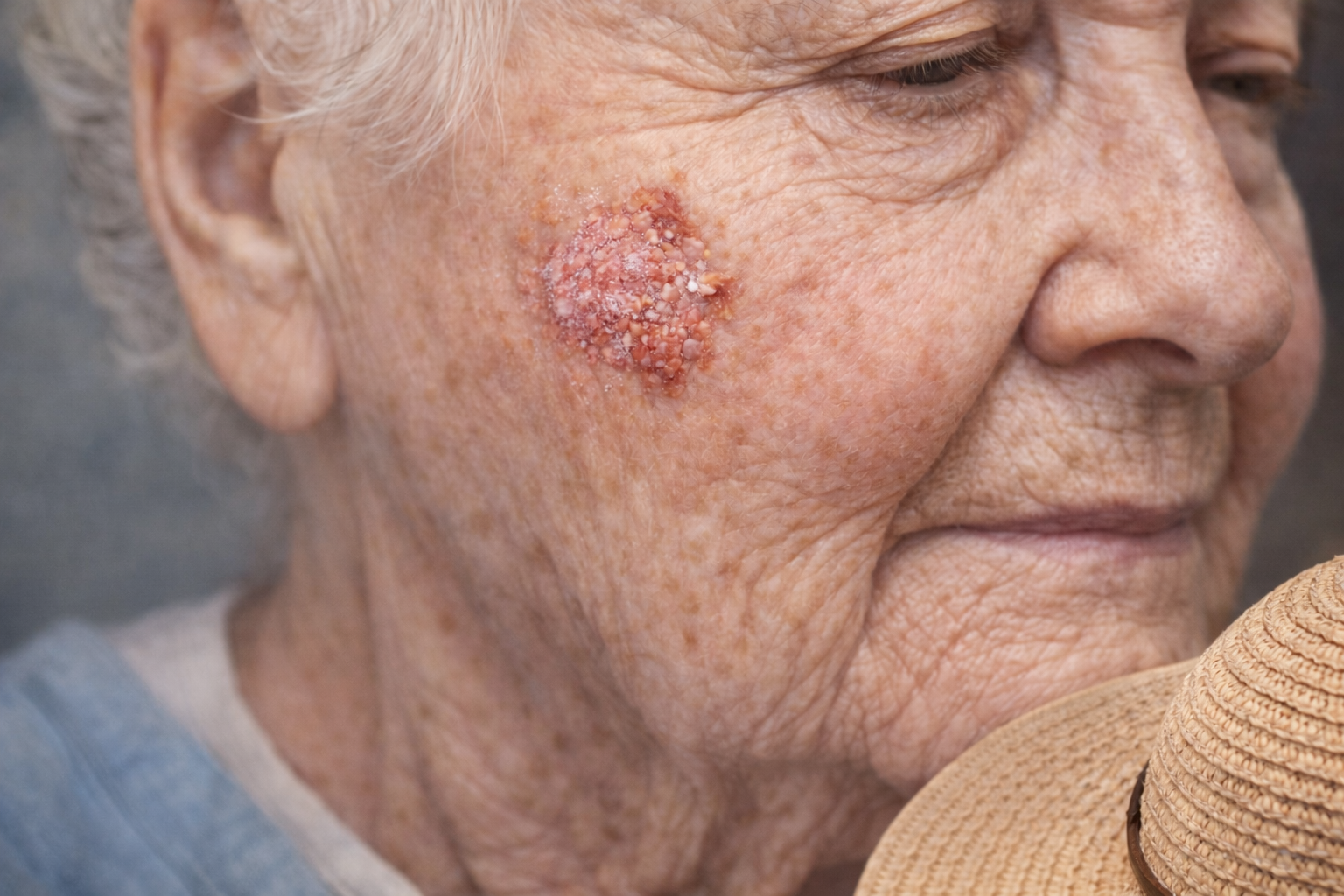
Actinic Keratosis Case Report: Treating a Precancerous Facial Lesion in a 92-Year-Old Patient
A 92-year-old female with a precancerous facial lesion was treated with liquid nitrogen cryotherapy. Learn how Village Dermatology in Katy and Houston, Texas manages actinic keratosis to reduce skin cancer risk.
By: Dr. Ashley Baldree
At Village Dermatology, early detection and treatment of precancerous skin lesions is a critical part of caring for our aging population in Katy and Houston, Texas. Actinic keratoses (AKs) are common in older adults with cumulative sun exposure and require prompt evaluation to reduce the risk of progression to skin cancer.
Patient Presentation
A 92-year-old female presented as a new patient for evaluation of a changing skin lesion on the left cheek. The lesion had been present for several months and was described as darkening, enlarging, and irregular, raising concern for sun-related precancerous change. The lesion had not been previously treated.
The patient presented for full dermatologic evaluation and management.
Comprehensive Skin Examination
A full-body skin examination was performed, including the scalp, face, neck, trunk, upper and lower extremities, hands, and forearms. Dermoscopy was used to further evaluate the lesion.
The patient appeared well-developed, well-nourished, alert, and in no acute distress.
On examination, there was a hypertrophic erythematous papule with hyperkeratotic scale located on the left medial malar cheek, clinically consistent with actinic keratosis.
Assessment
Actinic Keratosis (AK) – L57.0
Precancerous lesion on sun-damaged facial skin
Treatment Plan
Given the clinical appearance and risk factors, liquid nitrogen cryotherapy was recommended and performed during the visit.
Cryotherapy Details
1 lesion treated
3 freeze–thaw cycles
Location: left medial malar cheek
Informed consent was obtained, including discussion of potential risks such as blistering, scabbing, pigmentary changes, scarring, infection, recurrence, and incomplete removal.
If the lesion does not fully resolve, shave biopsy may be considered at a future visit.
Patient Counseling & Education
The patient received thorough counseling regarding actinic keratoses, including:
Skin Cancer Prevention
Daily use of broad-spectrum sunscreen SPF 30+
Wearing sun-protective clothing
Avoiding peak sun exposure when possible
Expectations
Actinic keratoses are precancerous growths caused by long-term sun exposure
While many AKs respond well to treatment, a small percentage may progress to squamous cell carcinoma if left untreated
When to Contact the Office
If the lesion does not resolve
If new or changing lesions appear
If severe side effects occur, such as excessive crusting, tenderness, or redness
High-Quality Skin Cancer Prevention in Katy & Houston
This case highlights the importance of early recognition and treatment of precancerous lesions, especially in older adults. At Village Dermatology, we emphasize preventive care, patient education, and evidence-based treatments to help reduce the risk of skin cancer.
If you or a loved one has a new or changing skin lesion, our dermatology team is here to help.

Dog Bite to the Lip: Prompt Dermatologic Care in Katy & Houston, Texas
A 55-year-old woman presents to Village Dermatology in Katy and Houston, Texas with a dog bite to the lower lip, treated with antibiotics, topical therapy, and detailed wound care counseling.
Dr. Ashley Baldree
Case Overview
A 55-year-old female new patient presented to Village Dermatology after sustaining a dog bite to the right lower lip earlier the same morning. She reported pain, redness, and moderate severity at the site of injury. Facial dog bites require prompt evaluation due to the risk of infection, scarring, and involvement of sensitive structures.
Clinical Examination
A focused examination of the head and face was performed using dermoscopy. The patient was well developed, well nourished, alert, oriented, and in no acute distress. Examination revealed puncture wounds on the right inferior vermilion lip, consistent with a recent dog bite. No signs of systemic infection were present at the time of evaluation.
Diagnosis: Dog Bite (Initial Encounter)
The patient was diagnosed with a dog bite to the lower lip, a common but potentially serious type of animal bite. Facial dog bites are carefully managed due to higher infection risk and cosmetic concerns.
Treatment Plan for Dog Bite Management
The patient was counseled extensively on wound care and infection prevention. Key components of the treatment plan included:
Infection Prevention
Oral antibiotics: Amoxicillin-clavulanate prescribed twice daily for 10 days
Topical antibiotic: Mupirocin ointment applied twice daily until healed
Dog bites are not sutured due to increased infection risk, especially when puncture wounds are present.
Wound Care Instructions
Clean the wound thoroughly with soap and water
Perform vinegar soaks (1:1 ratio) for 5 minutes, up to three times daily
Apply mupirocin ointment after soaks
Monitor closely for signs of infection
The patient was instructed to seek emergency care if she develops fever, chills, increasing redness, swelling, or worsening pain.
Scar Prevention Counseling
The importance of meticulous wound care to minimize scarring was emphasized. The patient was advised to allow complete healing before pursuing scar treatments. Silicone-based scar therapy will be discussed at follow-up.
Additional Counseling for Animal Bites
The patient was educated on:
The importance of identifying the animal involved
Possible need for rabies evaluation if the animal cannot be observed
Tetanus vaccination considerations
When emergency department evaluation is necessary
Additional Finding: Verruca Vulgaris
During the visit, verruca vulgaris (common warts) were also noted on the left distal dorsal forearm. The patient was counseled on treatment options including topical therapies and cryotherapy, with expectations for resolution discussed.
Expert Dermatologic Care for Animal Bites in Katy & Houston
At Village Dermatology, we provide prompt evaluation and treatment of dog bites and facial wounds, focusing on infection prevention, proper healing, and minimizing long-term scarring. Our dermatologists serve patients throughout Katy and Houston, Texas, offering expert care for both urgent and routine skin concerns.
If you experience an animal bite or facial injury, early dermatologic evaluation is essential for the best outcome.
Facial Discoloration and Textured Skin: Treating Irritant Contact Dermatitis in Katy & Houston, Texas
A 30-year-old woman presents to Village Dermatology in Katy and Houston, Texas with facial discoloration and textured skin diagnosed as irritant contact dermatitis with post-inflammatory hyperpigmentation.
By: Dr. Caroline Vaughn
Case Overview
A 30-year-old female new patient presented to Village Dermatology with concerns of facial discoloration affecting both cheeks. She also noted skin texture changes and intermittent breakouts. The patient had a prior history of completing an isotretinoin (Accutane) course and reported that her typical acne had remained well controlled since then.
Clinical Examination
A comprehensive facial examination was performed, including dermoscopic evaluation and palpation of the supraclavicular lymph nodes. The patient appeared well developed, well nourished, alert, oriented, and in no acute distress. Examination findings were consistent with irritant contact dermatitis, with associated post-inflammatory discoloration.
Diagnosis: Irritant Contact Dermatitis
Based on clinical findings, the discoloration and rough skin texture were attributed to irritant contact dermatitis (ICD). The patient reported using multiple over-the-counter serums and an exfoliator, which likely contributed to skin barrier disruption and irritation.
Patients with ICD often develop redness, texture changes, and discoloration when the skin is exposed to harsh or excessive skincare products. In this case, the discoloration was explained as post-inflammatory hyperpigmentation resulting from the underlying rash.
Treatment Plan: Simplifying Skincare
The patient was counseled extensively on simplifying her skincare routine to allow the skin barrier to heal. Key recommendations included:
Gentle Skincare Routine
Avoid harsh chemicals, exfoliators, and overuse of active ingredients
Use gentle cleansers and fragrance-free moisturizers
Apply moisturizers regularly to reduce irritation
Consider topical steroids if inflammation worsens
Patients were advised that irritant contact dermatitis may persist unless triggering products are eliminated, and patch testing may be considered if symptoms fail to improve.
Managing Hyperpigmentation
The patient was also diagnosed with post-inflammatory hyperpigmentation (PIH). Counseling emphasized that PIH may take months to years to fade but typically improves over time with proper care.
Hyperpigmentation Treatment Recommendations
Strict sun protection with broad-spectrum sunscreen SPF 30+
Protective clothing and minimizing sun exposure
Prescription hydroquinone applied nightly to affected areas for three months, followed by a one-month break before restarting if needed
Recommended Sunscreens for Sensitive Skin
To prevent worsening discoloration, the following facial sunscreens were recommended:
EltaMD UV Clear (Tinted)
Supergoop! Glowscreen
InnBeauty Mineral Glow Screen
ISDIN Eryfotona Actinica
La Roche-Posay Anthelios Face Sunscreen
Recommended Moisturizers
To support barrier repair and reduce irritation:
Vanicream Daily Facial Moisturizer
CeraVe PM Facial Moisturizing Lotion
La Roche-Posay Toleriane Double Repair
Avène Cicalfate
Kiehl’s Ultra Facial Cream
Expert Dermatologic Care in Katy & Houston
At Village Dermatology, we specialize in diagnosing and treating facial rashes, discoloration, and skin texture changes. Whether your concerns stem from sensitive skin, product reactions, or post-inflammatory hyperpigmentation, our dermatologists provide personalized treatment plans to restore healthy, even-toned skin.
If you’re experiencing persistent facial discoloration or irritation, schedule an evaluation with Village Dermatology in Katy or Houston, Texas for expert care.

Aging Face and Sun Spots: A Personalized Anti-Aging Consultation in Katy & Houston, Texas
A 42-year-old woman visits Village Dermatology in Katy and Houston, Texas to address facial aging and sun spots, exploring tretinoin, microneedling, PRP, laser resurfacing, and personalized anti-aging skincare options.
By: Dr. Caroline Vaughn
Case Overview
A 42-year-old female established patient presented to Village Dermatology for evaluation of facial aging and sun spots. Her primary concerns included changes in skin texture, uneven tone, and visible signs of sun damage. She was interested in learning about effective anti-aging skincare products and cosmetic procedures to improve her overall appearance while maintaining natural-looking results.
Clinical Examination
A focused facial examination was performed. The patient appeared well developed and well nourished, alert, oriented, and in no acute distress. Findings were consistent with age-related skin texture changes and sun-related pigmentation, common concerns among patients seeking cosmetic dermatology care in Katy and Houston, Texas.
Treatment Discussion: Anti-Aging Options
During the visit, a comprehensive discussion was held regarding both topical and procedural anti-aging treatments:
Medical-Grade Topical Therapy
The patient was counseled on the benefits of topical retinoids for improving skin texture, fine lines, and sun damage:
Tretinoin 0.025% cream was prescribed to be applied nightly as tolerated using a pea-sized amount for the entire face.
If irritation occurs, the patient may transition to over-the-counter Differin (adapalene) as a gentler alternative.
Retinoids remain a cornerstone of anti-aging skincare, stimulating collagen production and promoting smoother, more even-toned skin.
Procedural Anti-Aging Options
Several in-office cosmetic procedures were discussed to address varying levels of skin aging:
CO₂ Laser Resurfacing for more aggressive treatment of wrinkles, sun spots, and texture irregularities.
Microneedling as a less aggressive option to improve collagen production and skin tone.
Microneedling with PRP (Platelet-Rich Plasma) to enhance rejuvenation and healing.
Dermal fillers to address under-eye volume loss and restore a refreshed appearance.
A pricing sheet was provided, allowing the patient to review options at her convenience.
Sun Protection & Skincare Counseling
Given the role of sun exposure in premature aging, the patient was counseled extensively on daily sun protection. Recommended facial sunscreens included:
EltaMD UV Clear (Tinted)
Supergoop! Glowscreen
InnBeauty Mineral Glow Screen
ISDIN Eryfotona Actinica
La Roche-Posay Anthelios Face Sunscreen
Daily broad-spectrum sunscreen use is essential for preventing further sun damage and maintaining results from anti-aging treatments.
Additional Consideration: Rosacea
The patient also has a history of rosacea. Laser treatment options were discussed for redness and visible blood vessels; however, she elected to defer treatment at this time. Counseling emphasized:
Regular sunscreen use
Trigger avoidance (heat, spicy foods, alcohol, stress)
Understanding that rosacea is a chronic condition that can be managed with appropriate care
Personalized Cosmetic Dermatology in Katy & Houston
At Village Dermatology, we offer individualized anti-aging and cosmetic dermatology solutions designed to address sun damage, skin texture changes, and facial aging. From prescription skincare to advanced laser treatments, our team helps patients in Katy and Houston, Texas achieve healthy, youthful-looking skin with confidence.
If you’re noticing sun spots, fine lines, or changes in skin texture, schedule a cosmetic consultation to explore the best options for your skin.
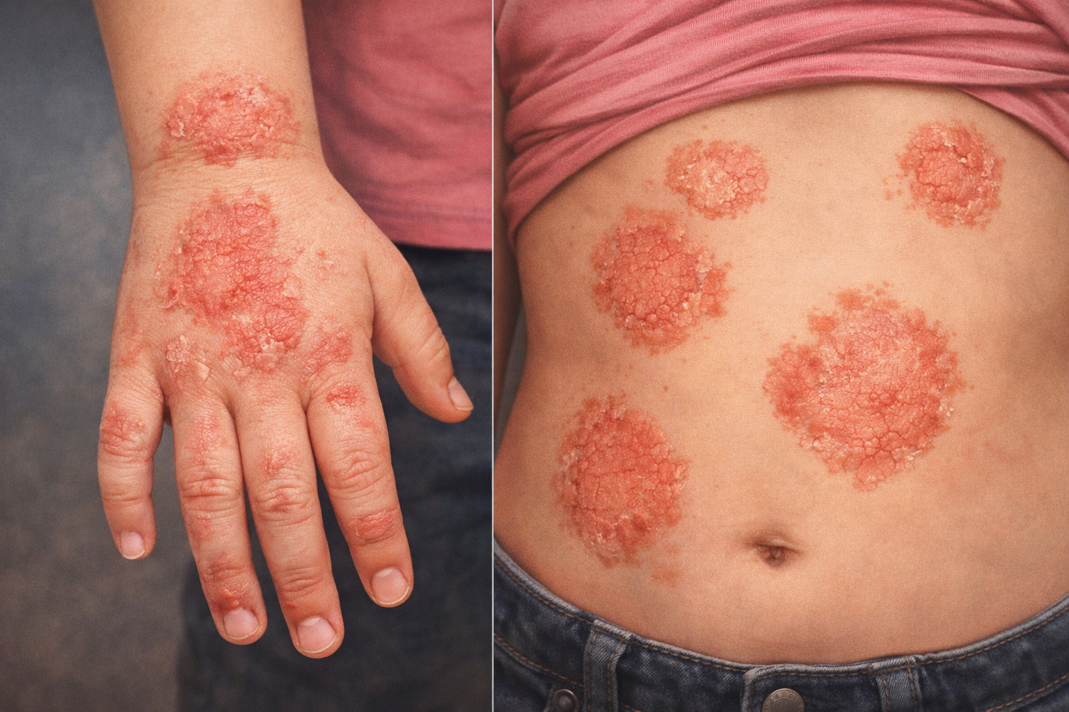
Pediatric Wart Removal Case: Treating Verruca Vulgaris in a 12-Year-Old Patient
A 12-year-old female with persistent warts on the knee was successfully treated with liquid nitrogen cryotherapy. Learn how Village Dermatology in Katy and Houston, Texas manages pediatric verruca vulgaris safely and effectively.
At Village Dermatology, we commonly treat viral warts (verruca vulgaris) in children and adolescents. Warts are benign but often persistent skin growths caused by the human papillomavirus (HPV) and may spread with contact or minor skin trauma. This case highlights effective in-office treatment and at-home management for pediatric warts in Katy and Houston, Texas.
Patient Presentation
A 12-year-old female presented as a new patient for evaluation of a flat wart on the right knee that had been present for several months. The lesion was first noticed by her mother during a field hockey tournament and persisted despite observation. The patient was referred by her pediatrician for dermatologic evaluation and possible removal.
Clinical Examination
A focused examination of the right lower extremity was performed using dermoscopy. The patient appeared well-developed, well-nourished, and in no acute distress.
On exam, there were two pink, cauliflower-like papules consistent with verruca vulgaris located on:
Right knee
Right proximal pretibial region
Right medial proximal pretibial region
Assessment
Treatment Plan
After reviewing the diagnosis, etiology, and treatment options with the patient and her mother, liquid nitrogen cryotherapy was recommended and performed during the visit.
Cryotherapy Details:
2 lesions treated
2 freeze–thaw cycles per lesion
Locations: right knee and right medial proximal pretibial region
Informed consent was obtained, including discussion of possible side effects such as blistering, scabbing, pigmentary changes, scarring, recurrence, incomplete removal, and infection.
At-Home Wart Care & Counseling
The patient and her mother were counseled extensively on wart management and prevention:
Treatment Options
Cryotherapy
Salicylic acid preparations
Retinoids
Aldara® (imiquimod), when appropriate
Home Care Instructions
Apply over-the-counter maximum strength salicylic acid bandages nightly for two weeks between monthly visits
This helps reduce wart size and may decrease the need for repeated in-office treatments
Education & Expectations
Warts are caused by a viral infection
They can spread through direct skin contact
With consistent treatment, most warts resolve successfully
Patients were advised to contact the office if:
Warts spread
Lesions recur
There is no improvement with treatment
Follow-Up
Return visit scheduled in 4 weeks for reassessment and possible repeat treatment
Expert Pediatric Wart Treatment in Katy & Houston
This case demonstrates the importance of early treatment and patient education when managing pediatric warts. At Village Dermatology, we offer safe, effective wart removal for children and teens using evidence-based therapies in a comfortable, family-friendly setting.
If your child has persistent warts or other skin concerns, our dermatology team is here to help.
Pediatric Eczema Follow-Up Case: Improving Chronic Atopic Dermatitis in a 5-Year-Old Patient
A 5-year-old female with chronic eczema shows improvement with topical therapy but continues to experience flares. Learn how Village Dermatology in Katy and Houston, Texas optimizes pediatric eczema care with advanced treatments and family education.
At Village Dermatology, we frequently care for children with eczema (atopic dermatitis), a chronic but manageable skin condition that can significantly affect quality of life for both patients and their families. This case highlights the importance of consistent skin care, medication optimization, and long-term management for pediatric eczema patients in Katy and Houston, Texas.
Patient Presentation
A 5-year-old female returned to our clinic for a follow-up evaluation of eczema affecting the right hand and trunk. She was initially seen in August 2024 and started on triamcinolone acetonide 0.1% topical cream, applied twice daily during flares with a maximum of 14 days per month.
At this visit, her father reported overall improvement with treatment; however, the child continued to experience intermittent flares and nighttime itching, prompting further evaluation and adjustment of her treatment plan.
Clinical Examination
A focused skin examination was performed, including the chest, bilateral forearms, and lower legs. Dermoscopic evaluation revealed coin-shaped (nummular) eczematous patches on the right hand and trunk. The patient appeared well-developed, well-nourished, and in no acute distress.
Assessment
Atopic dermatitis (eczema), chronic with flares
Coin-like eczematous patches on the right hand and trunk
Treatment Plan & Counseling
Given the persistent flares, the treatment plan was optimized to improve symptom control while minimizing long-term steroid exposure:
Continue triamcinolone acetonide 0.1% cream for flares (refill provided)
Initiate Vtama® (tapinarof) 1% topical cream, applied once daily to affected areas
Start cetirizine (Zyrtec®) at night to help reduce itching and improve sleep
Follow-up scheduled in 2 months
Extensive counseling was provided to the patient’s family, emphasizing:
Skin Care Routine
Bathe with lukewarm water using a gentle, fragrance-free cleanser
Apply moisturizer immediately after bathing
Use thick emollients 2–3 times daily
Avoid scented detergents, soaps, and fabric softeners
Expectations & Education
Families were counseled that eczema is chronic and relapsing, often triggered by:
Dry skin
Weather changes
Scratching
Stress
Scented products
Skin infections
Parents were advised to contact our office if symptoms worsen, fail to improve, or if signs of infection such as yellow crusting or painful sores develop.
Medication counseling included discussion of:
Possible mild burning with topical non-steroidal treatments
Safe use of topical steroids and avoidance of high-potency steroids on the face, groin, or skin folds
Potential side effects of prolonged steroid use, including skin thinning and discoloration
All questions were addressed, and the family demonstrated understanding of the treatment plan.
Comprehensive Pediatric Eczema Care in Katy & Houston
This case underscores the importance of personalized eczema management for children. At Village Dermatology, we offer expert care using the latest treatments—including steroid-sparing topical therapies—to help children achieve long-term skin comfort and healthier skin.
If your child is struggling with eczema or recurrent rashes, our board-certified dermatology team is here to help.
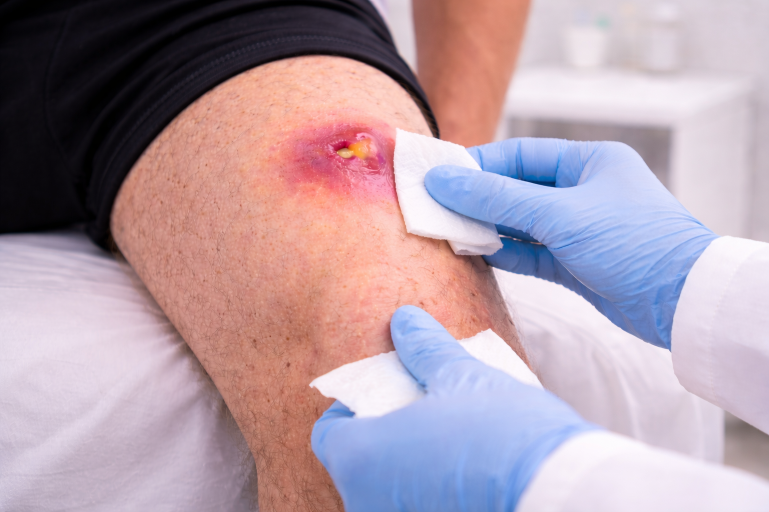
Painful Skin Abscess Treated With Incision and Drainage in a 28-Year-Old Male
A 28-year-old male with a painful skin abscess underwent incision and drainage at Village Dermatology in Katy and Houston, Texas, highlighting expert infection management and procedural dermatologic care.
Village Dermatology | Katy & Houston, Texas
Overview
A 28-year-old male presented to Village Dermatology with a painful skin infection on the back of his left thigh. The area had become increasingly tender and inflamed over the course of several days despite oral antibiotic therapy, prompting further evaluation and treatment.
This case highlights the importance of timely incision and drainage (I&D) for skin abscesses that do not adequately respond to antibiotics alone.
Clinical Evaluation
Focused examination of the left posterior thigh revealed a localized, inflamed lesion consistent with a cutaneous abscess. The patient was otherwise healthy, well-appearing, and in no acute distress.
Findings were concerning for a collection of pus beneath the skin, a condition that often requires procedural intervention to achieve full resolution.
Diagnosis: Skin Abscess
The lesion was diagnosed as a skin abscess, a localized bacterial infection that forms a pocket of purulent material. Abscesses commonly cause:
Pain and tenderness
Redness and warmth
Swelling and pressure
Because abscesses can be caused by bacteria such as Staphylococcus aureus, including MRSA, culture and drainage are often necessary for effective treatment.
Treatment Plan
Given the patient’s persistent pain and incomplete response to antibiotics, incision and drainage was recommended and performed during the visit.
Treatment included:
Incision and drainage of the abscess under local anesthesia
Expression of purulent contents
Application of a pressure dressing
Bacterial culture to guide ongoing treatment
The procedure was medically necessary due to infection, pain, redness, and failure of conservative measures.
Post-Procedure Care & Counseling
The patient was counseled on:
Proper wound care and dressing changes
Use of antibacterial cleansers such as benzoyl peroxide or chlorhexidine to reduce bacterial load
The importance of completing prescribed antibiotics
Monitoring for warning signs such as fever, chills, worsening pain, or spreading redness
Warm compresses were also recommended to promote continued drainage and healing.
Follow-Up Plan
The patient was instructed to follow up if:
The abscess does not improve
Symptoms worsen
Systemic signs of infection develop
Prompt follow-up ensures proper healing and reduces the risk of recurrence.
Expert Abscess Treatment in Katy & Houston
Skin abscesses can become serious if not treated appropriately. At Village Dermatology, we provide same-day evaluation and procedural care, including incision and drainage, to relieve pain and prevent complications.
If you have a painful, swollen skin infection, our dermatology team in Katy and Houston, Texas is here to help.
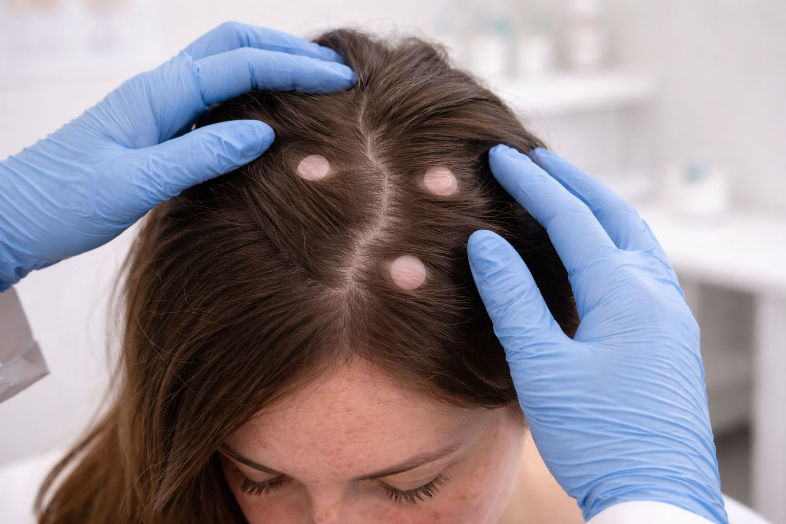
Multiple Pilar Cysts of the Scalp in a 36-Year-Old Female
A 36-year-old female with multiple pilar cysts of the scalp was evaluated at Village Dermatology in Katy and Houston, Texas and scheduled for in-office cyst excision.
Village Dermatology | Katy & Houston, Texas
Overview
A 36-year-old female returned to Village Dermatology with concerns about several firm bumps on her scalp that had slowly developed over the past few months. She reported a history of similar scalp cysts that were previously removed, prompting evaluation to confirm the diagnosis and discuss management options.
This case highlights the diagnosis and treatment planning for pilar cysts, a common benign scalp condition.
Clinical Evaluation
A focused scalp examination was performed with dermoscopic evaluation. The patient appeared healthy, well-nourished, and in no acute distress.
On examination, three discrete, firm, subcutaneous nodules were identified on the scalp. These lesions were smooth, non-inflamed, and consistent with benign cysts arising from the hair follicle.
Diagnosis: Pilar Cysts
Based on clinical findings and patient history, the lesions were diagnosed as pilar cysts—benign keratin-filled cysts that commonly occur on the scalp and may be multiple or recurrent. Pilar cysts often have a genetic predisposition and are more frequently seen in women.
The cysts measured approximately:
0.5 cm on the right frontal scalp
0.7 cm on the left frontal scalp
0.6 cm on the left parietal scalp
Treatment Plan
The benign nature of pilar cysts was reviewed in detail. Because the lesions were bothersome and the patient had a prior history of cyst excisions, she elected to proceed with surgical removal.
Management plan included:
Scheduling an in-office cyst excision
Counseling on the minor surgical procedure, healing expectations, and scarring
Reassurance that no specific skincare or medical treatment is required prior to excision
Patient Education & Counseling
The patient was counseled that:
Pilar cysts are non-cancerous
They may slowly enlarge over time
Treatment is optional unless cysts become painful, inflamed, or cosmetically bothersome
She was advised to contact the office if any cyst becomes red, tender, or ruptures, which may indicate inflammation or infection.
Expert Scalp Care in Katy & Houston
Scalp cysts are a common concern and can often recur without proper evaluation. At Village Dermatology, we offer expert diagnosis and in-office surgical treatment for benign scalp lesions in a safe, comfortable setting.
If you have bumps on the scalp or recurrent cysts, our dermatology team in Katy and Houston, Texas is here to help.