
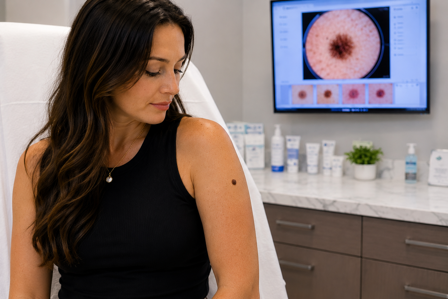
"Doctor, Should I Be Worried About This Mole on My Arm?"
A 31-year-old Texas patient underwent a full body skin examination after concerns about a mole on her arm. Learn how dermatologists evaluate moles, identify benign skin growths, and detect skin cancer early at Village Dermatology in Katy and Houston, Texas.
A Full Body Skin Exam Reveals Common Benign Skin Growths in a Young Texas Patient
At Village Dermatology, one of the most common questions we hear from patients in Katy and Houston is:
"Doctor, should I be worried about this mole on my arm?"
A recent patient visit highlights why routine skin examinations are an important part of maintaining healthy skin and why many seemingly concerning lesions turn out to be completely benign.
Patient Case Overview
A 31-year-old woman presented to Village Dermatology for her annual full body skin examination. She had several skin lesions she wanted evaluated, including spots on her left upper arm and left lower leg.
Although the lesions were not causing pain, itching, or bleeding, she wanted reassurance that they were not dangerous and sought a comprehensive skin cancer screening.
Comprehensive Skin Examination
A thorough full body skin examination was performed, including evaluation of:
Scalp and hair
Face and neck
Chest and abdomen
Back
Arms and legs
Hands and feet
Nails
Other skin surfaces
A dermatoscope was utilized to closely examine pigmented lesions and identify features that may indicate skin cancer or other concerning changes.
Findings: Benign Nevi (Moles)
The primary lesion of concern on the left upper arm measured approximately 6.5 mm and demonstrated reassuring features:
Symmetrical appearance
Regular borders
Even pigmentation
Stable clinical appearance
The lesion was diagnosed as a benign nevus (common mole).
Benign nevi are collections of pigment-producing cells called melanocytes. Most adults develop multiple moles throughout their lifetime, and the vast majority are harmless.
Additional Benign Findings
During the examination, several other common benign skin conditions were identified.
Seborrheic Keratoses
A waxy, pigmented growth on the trunk was diagnosed as a seborrheic keratosis.
These growths are:
Extremely common
Non-cancerous
Often hereditary
More common with age
Many patients mistake seborrheic keratoses for skin cancer because they can appear dark, raised, and irregular.
Lentigines (Sun Spots)
The patient also demonstrated lentigines, commonly called:
Age spots
Sun spots
Liver spots
These develop due to cumulative ultraviolet (UV) exposure and are especially common in sunny climates like Katy and Houston.
While benign, they serve as a reminder that the skin has experienced sun damage over time.
Cherry Angiomas
Multiple cherry angiomas were noted during the examination.
These bright red vascular growths are:
Benign
Extremely common
Typically increase with age
Not associated with skin cancer
Dermatofibroma
A dermatofibroma was identified on the left upper back.
Dermatofibromas are firm, scar-like nodules that often develop following minor trauma such as:
Insect bites
Ingrown hairs
Small cuts or scratches
These lesions are benign and usually require no treatment unless they become symptomatic.
Changes in Skin Texture
The patient also had an area of altered skin texture on the forehead. While no definitive diagnosis was established during the visit, conservative management with moisturization and topical tretinoin therapy was initiated.
Close monitoring was recommended to ensure no progression occurs.
Why Annual Skin Exams Matter
Many patients assume they only need a skin examination if they have a suspicious lesion. However, routine skin checks provide several important benefits:
Early detection of melanoma
Identification of precancerous lesions
Monitoring of changing moles
Education regarding sun protection
Peace of mind regarding benign growths
Even younger adults can develop skin cancer, making periodic skin examinations an important preventive healthcare measure.
How to Monitor Moles at Home
Patients are encouraged to perform monthly self-skin examinations using the ABCDE rule:
A – Asymmetry
One half differs from the other.
B – Border
Edges are irregular or poorly defined.
C – Color
Multiple colors or uneven pigmentation.
D – Diameter
Larger than 6 mm or increasing in size.
E – Evolving
Any change in appearance, symptoms, or behavior.
If a mole changes in size, shape, color, begins itching, bleeding, or becomes painful, evaluation by a board-certified dermatologist is recommended.
Protecting Your Skin in Texas
Residents of Katy and Houston experience significant UV exposure throughout the year. Consistent sun protection remains one of the most effective ways to prevent skin cancer and premature aging.
Recommended sun safety measures include:
Daily broad-spectrum SPF 30 or higher sunscreen
Reapplication every two hours outdoors
Sun-protective clothing
Wide-brimmed hats
UV-blocking sunglasses
Seeking shade during peak sunlight hours
Expert Skin Cancer Screening in Katy and Houston
At Village Dermatology, we provide comprehensive skin cancer screenings, mole evaluations, and personalized dermatologic care for patients throughout Katy, Houston, and surrounding communities.
If you have a mole or skin lesion that concerns you, scheduling a professional skin examination can provide reassurance and help identify potential problems early.
Our goal is to help every patient maintain healthy skin through prevention, education, and expert dermatologic care.
Treating Stubborn Plantar Warts in Children: A Follow-Up Success Story
A young patient returned for follow-up treatment of a stubborn plantar wart on the foot. Learn how cryotherapy and at-home salicylic acid therapy help eliminate persistent warts in children.
Pediatric Wart Treatment in Katy and Houston, Texas
Plantar warts are a common skin condition in children and can often be frustrating for both patients and parents. These warts develop on the feet and are caused by infection with the human papillomavirus (HPV). While many warts eventually resolve on their own, treatment is often recommended when they become persistent, uncomfortable, or continue to spread.
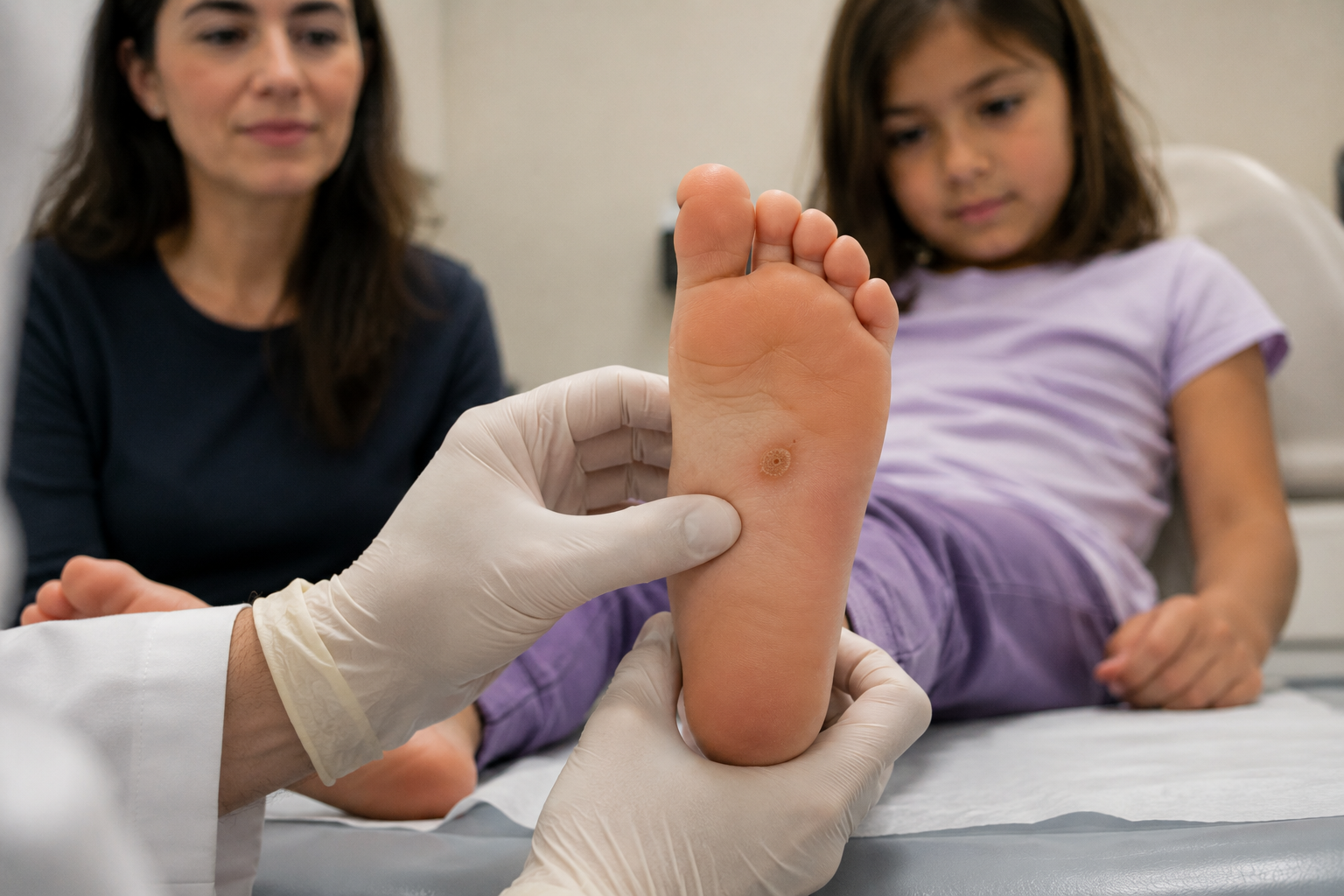
At Village Dermatology, we recently followed a young patient who returned for ongoing treatment of a plantar wart located on the arch of her foot.
Patient Presentation
A 7-year-old female returned for follow-up evaluation of a verruca vulgaris (common wart) located on the right arch of her foot.
At her previous visit, treatment included:
Careful paring of the wart with a surgical blade
Liquid nitrogen cryotherapy (LN2)
Instructions to begin over-the-counter salicylic acid wart bandages at home
The patient and her mother returned for reevaluation to assess treatment progress.
Why Plantar Warts Can Be Difficult to Treat
Plantar warts develop on weight-bearing areas of the foot, making them particularly stubborn.
Factors that contribute to persistent plantar warts include:
Pressure from walking and standing
Thick skin on the soles of the feet
Deep extension of wart tissue
Ongoing viral activity within the skin
Because of these factors, multiple treatment sessions are often necessary to achieve complete clearance.
Follow-Up Examination
During the visit, a comprehensive examination of the patient's right foot was performed.
Although the wart had been previously treated, examination revealed that the lesion remained present on the right arch.
The patient and her mother discussed available treatment options, including:
Repeat Cryotherapy (Liquid Nitrogen)
Cryotherapy freezes the wart tissue and stimulates the body's immune system to help eliminate the viral infection.
Cantharidin Therapy
Cantharidin is a blistering agent frequently used in pediatric dermatology to treat warts.
After reviewing both options, the patient and her mother elected to proceed with another round of liquid nitrogen treatment.
Cryotherapy Treatment
The wart was treated with liquid nitrogen during the visit.
Cryotherapy remains one of the most commonly used wart treatments because it:
Is performed quickly in the office
Does not require surgery
Can be highly effective with repeated treatments
Helps stimulate an immune response against the wart virus
Patients and parents are counseled regarding possible temporary side effects including:
Blistering
Crusting
Scabbing
Temporary skin discoloration
Mild discomfort
These reactions are expected and often indicate that the treatment is working.
Home Wart Treatment Remains Important
Successful wart treatment often requires a combination of office-based therapy and at-home care.
Salicylic acid remains one of the most effective over-the-counter wart treatments and works by gradually removing infected skin cells.
Parents are encouraged to follow treatment instructions carefully and continue home therapy as recommended by their dermatologist.
Understanding Verruca Vulgaris
Verruca vulgaris, or common warts, are caused by HPV infection of the skin.
These lesions often appear as:
Rough or cauliflower-like bumps
Thickened areas of skin
Small flesh-colored or tan growths
Lesions that interrupt normal skin lines
Warts are contagious and may spread through direct contact or contact with contaminated surfaces.
Preventing Wart Spread
To help reduce transmission and recurrence, patients should:
Avoid picking at warts
Wear footwear in public locker rooms and pools
Avoid sharing shoes or socks
Keep affected areas clean and covered when appropriate
Follow all treatment recommendations
Early treatment can often prevent warts from becoming larger or spreading to additional locations.
Expert Pediatric Wart Treatment in Katy and Houston
At Village Dermatology, we provide comprehensive wart treatment for children and adults throughout Katy, Houston, and surrounding Texas communities. Treatment options include cryotherapy, salicylic acid therapy, cantharidin, and other advanced wart management techniques tailored to each patient.
If your child has a persistent wart that is not improving, schedule an appointment with our dermatology team for expert evaluation and treatment.
Enlarging Scalp Lesions: When Should You Have a Mole Checked?
Concerned about a scalp mole or enlarging skin lesion? Learn how Village Dermatology evaluated benign scalp nevi and a milia cyst in a young adult patient and when you should seek professional evaluation.
Scalp Mole Evaluation in Katy and Houston, Texas
Many people discover bumps or moles on their scalp while brushing their hair, visiting a hairstylist, or experiencing changes in existing skin lesions. Because the scalp is difficult to examine regularly, changes may go unnoticed for months or even years.
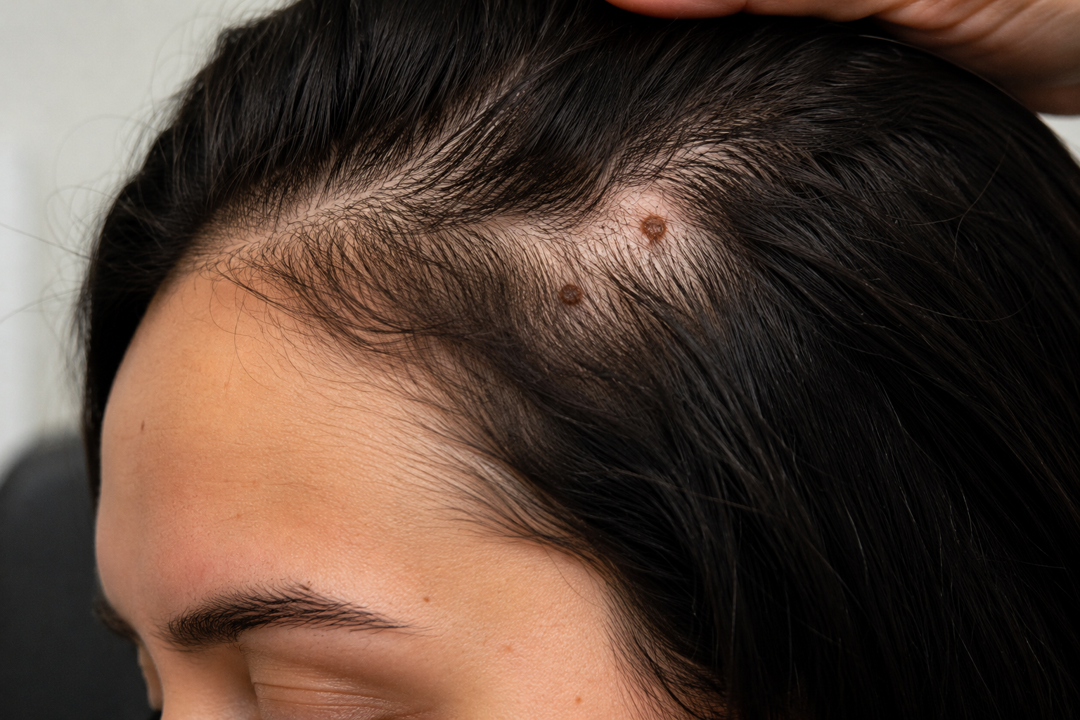
At Village Dermatology, we recently evaluated a young woman concerned about several enlarging lesions on her scalp that had been present for approximately two years.
Patient Presentation
A 25-year-old female presented as a new patient for evaluation of multiple scalp lesions. She reported:
Lesions located on the scalp
Gradual enlargement over time
No pain or itching
Presence for approximately two years
Concern regarding whether the growths were dangerous
Because changes in skin lesions can occasionally signal skin cancer or atypical moles, a comprehensive dermatologic evaluation was performed.
Comprehensive Scalp Examination
A detailed scalp examination was completed, including hair inspection and dermoscopic evaluation.
Dermoscopy is a specialized diagnostic technique that allows dermatologists to closely examine skin lesions using magnification and polarized light. This tool helps distinguish benign lesions from growths that may require further investigation.
Upon examination, the lesions demonstrated reassuring features including:
Symmetrical appearance
Uniform coloration
Regular borders
Stable pigmentation patterns
These findings were most consistent with benign melanocytic nevi, commonly known as moles.
Understanding Benign Scalp Moles
Benign nevi are collections of pigment-producing cells within the skin. They are extremely common and can occur anywhere on the body, including the scalp.
Many scalp moles remain stable throughout life and require no treatment.
Typical characteristics of benign moles include:
Symmetry
Even color distribution
Smooth borders
Stable appearance over time
Although most scalp moles are harmless, periodic monitoring remains important because changes can occur over time.
Why Scalp Moles Deserve Special Attention
The scalp is one of the most difficult areas for patients to monitor.
Hair often conceals lesions, making it challenging to identify changes in:
Size
Shape
Color
Surface texture
Because scalp melanomas can sometimes be diagnosed later than lesions found on other parts of the body, routine skin examinations are particularly valuable.
Patients should seek dermatologic evaluation if a scalp lesion:
Rapidly enlarges
Changes color
Becomes irregular
Bleeds
Itches persistently
Develops ulceration
Courtesy Extraction of a Milia Cyst
During the same visit, a small milia cyst was identified on the right central frontal scalp.
Milia are tiny superficial cysts that contain keratin, a naturally occurring skin protein. These small white or flesh-colored bumps are completely benign and frequently occur on the face and scalp.
The patient elected to undergo a courtesy extraction during the visit.
What Causes Milia?
Milia form when keratin becomes trapped beneath the skin surface.
Common causes include:
Natural skin cell turnover
Skin trauma
Sun damage
Occlusive skincare products
Spontaneous development without a clear cause
Because milia are harmless, treatment is usually performed only for cosmetic reasons.
How to Monitor Moles at Home
Patients with moles should perform monthly self-skin examinations whenever possible.
The ABCDE warning signs can help identify concerning changes:
A – Asymmetry
One half differs from the other.
B – Border
Edges become irregular or poorly defined.
C – Color
Multiple colors develop within the lesion.
D – Diameter
Growth larger than approximately 6 mm.
E – Evolving
Any change in appearance over time.
If any of these changes occur, prompt dermatologic evaluation is recommended.
Expert Skin Cancer Screening and Mole Evaluation in Katy and Houston
At Village Dermatology, we provide comprehensive skin cancer screenings, mole evaluations, scalp examinations, and dermatologic surgery services for patients throughout Katy, Houston, and surrounding Texas communities.
If you notice a changing mole, enlarging scalp lesion, or any growth that concerns you, our dermatology team can provide an expert diagnosis and personalized treatment recommendations.
Teen Acne Success Story: Month 5 of Accutane Treatment Shows Excellent Progress
A 16-year-old acne patient completed month five of isotretinoin therapy with no new breakouts and minimal side effects. Learn how Accutane helps teens achieve long-term acne clearance.
Advanced Acne Treatment in Katy and Houston, Texas
Acne can be one of the most frustrating skin conditions for teenagers, especially when breakouts affect both the face and trunk. While many patients improve with topical medications and oral antibiotics, some cases require a more aggressive treatment approach to prevent scarring and achieve long-term clearance.
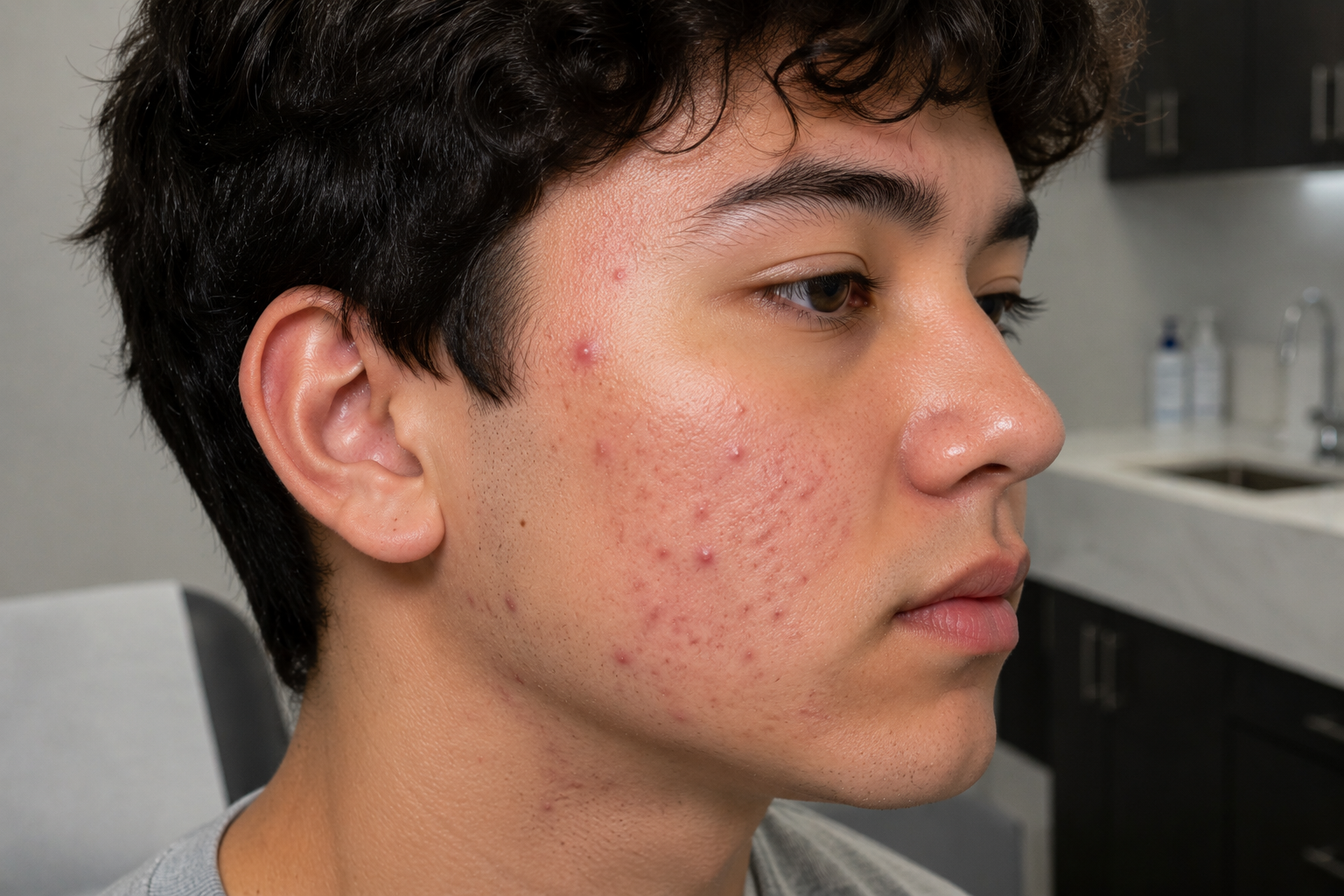
At Village Dermatology, we recently followed a 16-year-old patient undergoing isotretinoin (Accutane) therapy who demonstrated excellent progress during his fifth month of treatment.
Patient Background
The patient initially presented with inflammatory and comedonal acne involving the face and trunk. His acne included:
Inflammatory papules
Pustules
Comedonal lesions (clogged pores)
Persistent breakouts despite previous treatment attempts
Due to the severity and extent of his acne, isotretinoin therapy was initiated to target the underlying causes of acne and reduce the risk of permanent scarring.
Month 5 Follow-Up: Significant Improvement
During his recent follow-up appointment, the patient reported excellent tolerance of treatment.
Most importantly, he experienced:
No new acne breakouts during the previous month
Continued improvement of existing lesions
No significant treatment-related complications
Aside from manageable skin dryness, he reported no major side effects.
This type of response is encouraging and commonly seen as patients progress through later stages of isotretinoin therapy.
Isotretinoin Dosage Adjustment
Because the patient tolerated treatment well and continued to show improvement, his isotretinoin dosage was increased to 100 mg daily.
Dosage adjustments are often made throughout treatment based on:
Patient weight
Clinical response
Side effect profile
Cumulative target dose
The goal is to reach a cumulative treatment dose of approximately 200–220 mg/kg, which has been associated with lower relapse rates and long-term acne clearance.
Monitoring Safety During Accutane Therapy
One of the reasons isotretinoin remains such an effective treatment is that patients are closely monitored throughout therapy.
At this visit, the patient denied experiencing common isotretinoin side effects, including:
Depression or mood changes
Dry or cracked lips (cheilitis)
Severe skin dryness
Nosebleeds
Headaches
Muscle aches
Elevated cholesterol
Elevated triglycerides
Retinoid dermatitis
Because the dosage was increased, laboratory testing was scheduled for the following month to continue monitoring treatment safety.
Why Monthly Monitoring Matters
Isotretinoin is considered a high-risk medication that requires regular follow-up appointments and laboratory monitoring.
Patients receiving Accutane should:
Complete monthly evaluations
Undergo recommended blood testing
Avoid donating blood
Never share medication
Report any concerning symptoms immediately
Inform their dermatologist of any side effects
These precautions help ensure that treatment remains both safe and effective.
Skin Care During Accutane Treatment
Proper skincare is essential while taking isotretinoin.
Patients are encouraged to use:
Gentle Cleansers
Harsh scrubs and drying products should be avoided.
Non-Comedogenic Moisturizers
Regular moisturization helps minimize dryness and irritation.
Broad-Spectrum Sunscreen SPF 30+
Isotretinoin increases sun sensitivity, making daily sun protection extremely important.
Consistent skincare can significantly improve comfort throughout treatment.
What Results Can Patients Expect?
Many patients begin noticing substantial improvement after several months of therapy.
Isotretinoin works by addressing all major contributors to acne:
Excess oil production
Clogged pores
Inflammation
Acne-causing bacteria
For many patients, completing a full treatment course results in long-term remission and dramatically clearer skin.
Expert Teen Acne Treatment in Katy and Houston
At Village Dermatology, we specialize in personalized acne treatment plans for adolescents and adults throughout Katy, Houston, and surrounding communities.
Whether you are dealing with persistent acne, cystic acne, acne scarring, or treatment-resistant breakouts, our experienced dermatology team can help determine the most effective treatment strategy for your skin.
If you or your teenager are struggling with severe acne, schedule a consultation with Village Dermatology to learn whether isotretinoin may be an appropriate treatment option.
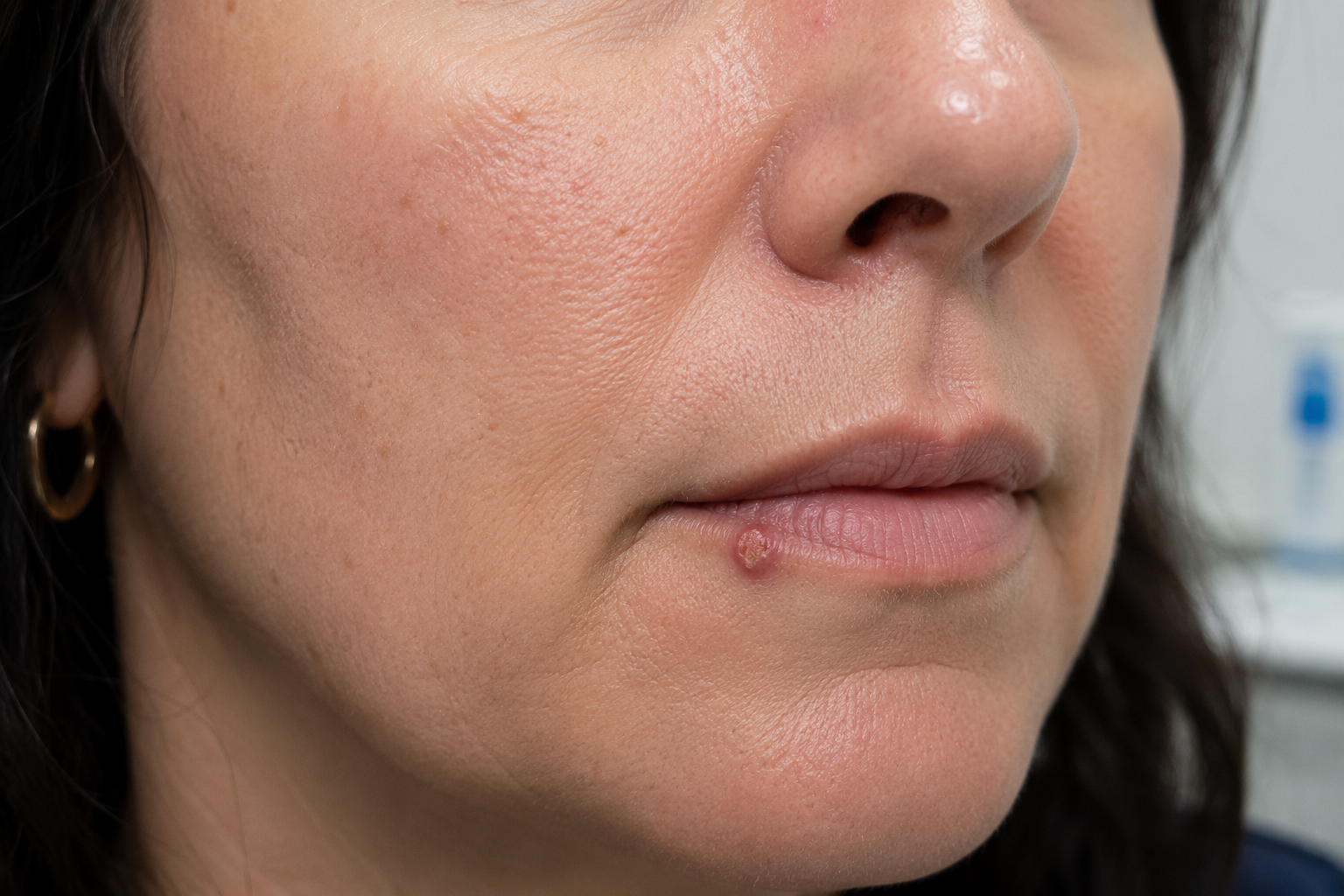
Wart on the Lip or Cold Sore? A Dermatologist Explains the Difference
Not every bump on the lip is a cold sore. Learn how Village Dermatology diagnosed and treated a persistent lip wart in a Houston-area patient and when a biopsy may be necessary.
by: Dr. Ashley Baldree
Evaluating a Persistent Lip Lesion in Katy and Houston, Texas
A new patient recently visited Village Dermatology concerned about a growth on her lower lip that had been present for approximately three months. The lesion first appeared shortly after a cold sore outbreak and had not resolved on its own.
Because growths on the lips can represent a variety of conditions—including warts, viral infections, benign growths, and, in rare cases, skin cancer—an accurate diagnosis by a board-certified dermatologist is important.
Patient Presentation
The patient was a 47-year-old woman who presented for evaluation of an asymptomatic lesion located on the right lower lip. She reported:
The lesion had been present for several months.
It developed after a cold sore outbreak.
The spot was not painful or itchy.
No prior treatment had been attempted.
No family history of melanoma.
A comprehensive skin examination of the face and lips was performed, including dermoscopic evaluation to closely examine the lesion's structure.
Examination Findings
Upon examination, a pink cauliflower-like papule was identified along the right inferior vermilion border of the lip.
The appearance was most consistent with verruca vulgaris, commonly known as a wart.
Warts are caused by infection with the human papillomavirus (HPV) and can occasionally develop on or around the lips. These lesions may appear rough, raised, or cauliflower-like and can persist for months if left untreated.
Because the lesion developed following a cold sore outbreak, it was understandable that the patient initially questioned whether the spot was related to herpes simplex virus. However, the clinical appearance suggested a wart rather than an active cold sore.
Treatment with Liquid Nitrogen Cryotherapy
After discussing treatment options, the patient elected to proceed with cryotherapy.
The lesion was carefully pared using a sterile blade before treatment. Liquid nitrogen was then applied using two freeze-thaw cycles to destroy the abnormal tissue.
Cryotherapy remains one of the most effective treatments for many common warts and offers several benefits:
Quick in-office procedure
Minimal downtime
No surgical incision required
High success rates for many wart types
Patients are counseled that temporary crusting, blistering, redness, or pigment changes can occur following treatment.
When Should a Lip Lesion Be Biopsied?
While many lip lesions are benign, persistent growths should never be ignored.
At Village Dermatology, we may recommend a biopsy when a lesion:
Does not respond to treatment
Continues to enlarge
Develops ulceration
Bleeds spontaneously
Has atypical clinical features
In this patient's case, a biopsy was discussed as a future option if the lesion fails to resolve after cryotherapy.
Managing Recurrent Cold Sores
During the visit, the patient also reported a history of recurrent cold sores.
Cold sores are caused by the herpes simplex virus (HSV-1) and commonly appear as painful clusters of blisters around the lips. While outbreaks often resolve on their own, many patients experience recurrent episodes triggered by:
Stress
Illness
Sun exposure
Trauma to the lips
Hormonal changes
For outbreak management, antiviral therapy was prescribed.
Preventing Future Herpes Simplex Outbreaks
Patients with recurrent cold sores can often reduce the severity and duration of outbreaks through early treatment and preventive measures.
Helpful strategies include:
Daily use of broad-spectrum SPF 30+ sunscreen
Avoiding excessive sun exposure
Managing stress levels
Beginning antiviral medication at the first sign of symptoms
Early intervention frequently shortens outbreaks and improves patient comfort.
Why Dermatology Evaluation Matters
Many patients assume any bump on the lip is a cold sore. However, dermatologists routinely diagnose a wide variety of lip lesions including:
Viral warts
Herpes simplex infections
Actinic cheilitis
Mucoceles
Benign growths
Precancerous lesions
Skin cancers
Obtaining an accurate diagnosis is essential for selecting the appropriate treatment and ensuring the best possible outcome.
Expert Wart and Cold Sore Treatment in Katy and Houston
At Village Dermatology, we diagnose and treat lip lesions, warts, cold sores, and skin cancers for patients throughout Katy, Houston, and surrounding communities.
If you have a persistent bump, wart, sore, or growth on your lip that does not heal, schedule an appointment with our dermatology team for a professional evaluation and personalized treatment plan.
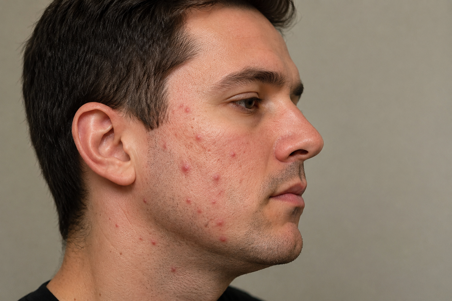
Acne Treatment Success with Isotretinoin (Accutane): Managing Side Effects While Achieving Clearer Skin
A 37-year-old patient undergoing Accutane treatment at Village Dermatology experienced improving acne with manageable side effects including dry lips and mild hair shedding. Learn how isotretinoin helps treat stubborn acne and what patients can expect during treatment.
by: Dr. Caroline Vaughn
Acne Treatment in Katy and Houston, Texas
Acne is one of the most common skin conditions treated at Village Dermatology. While many patients respond well to topical medications and oral antibiotics, some individuals require more advanced treatment to achieve lasting results. Isotretinoin, commonly known by its former brand name Accutane, remains one of the most effective treatments for moderate to severe acne.
Recently, we followed a 37-year-old male patient from the Katy and Houston area who was undergoing isotretinoin therapy for persistent facial acne.
The Patient's Acne Journey
The patient presented with moderate inflammatory and comedonal acne affecting both cheeks. His acne consisted of inflammatory papules, pustules, and clogged pores that had persisted for several months despite previous treatment attempts.
After a thorough evaluation, isotretinoin therapy was initiated to address the underlying causes of acne and reduce the risk of permanent scarring.
One-Month Follow-Up on Isotretinoin
At his one-month follow-up appointment, the patient reported that he was tolerating isotretinoin well overall. As expected, he experienced some common side effects including:
Dry lips (cheilitis)
Mild scalp hair shedding
Increased dryness of the skin
Fortunately, these side effects were manageable and did not interfere significantly with daily activities.
The patient had already been taking finasteride for androgenetic alopecia (male pattern hair loss). During the visit, we discussed that temporary increased hair shedding can occasionally occur while taking isotretinoin. In most cases, this side effect improves after treatment is completed.
Because the patient continued to experience a few new acne breakouts during the first month of therapy, his isotretinoin dosage was increased from 40 mg daily to 80 mg daily to help achieve optimal results.
Understanding Isotretinoin Side Effects
Many patients considering Accutane treatment are concerned about side effects. While isotretinoin is highly effective, close monitoring is essential.
Common side effects may include:
Dry lips
Dry skin
Dry eyes
Nosebleeds
Temporary hair shedding
Increased sun sensitivity
Patients receiving isotretinoin require regular follow-up appointments and laboratory monitoring throughout treatment.
At Village Dermatology, we carefully monitor all patients taking isotretinoin to ensure treatment remains both safe and effective.
Managing Cheilitis (Dry Lips) During Accutane Treatment
One of the most common side effects of isotretinoin is cheilitis, or inflammation and dryness of the lips.
To minimize discomfort, we recommend:
Frequent application of Vaseline® or Aquaphor®
Applying lip moisturizer before bedtime
Staying well hydrated
Avoiding lip licking, which can worsen irritation
Most patients find that consistent moisturization keeps symptoms manageable throughout treatment.
Important Safety Precautions for Accutane Patients
Patients taking isotretinoin should always follow safety guidelines, including:
Never sharing medication with others
Avoiding blood donation during treatment
Completing required monthly monitoring
Reporting any significant side effects immediately
Using sunscreen daily with SPF 30 or higher
Avoiding elective cosmetic procedures until advised by their dermatologist
Patient education is a critical part of successful isotretinoin therapy.
What Results Can Patients Expect?
Most patients begin seeing significant improvement after several months of treatment. While some breakouts may continue during the early stages, isotretinoin works by targeting all major causes of acne:
Excess oil production
Clogged pores
Inflammation
Acne-causing bacteria
Many patients experience long-term remission after completing a full cumulative treatment course.
Expert Acne Care in Katy and Houston
At Village Dermatology, we provide comprehensive acne treatment options for teenagers and adults throughout Katy, Houston, and surrounding communities. Whether you are struggling with persistent breakouts, acne scarring, or treatment-resistant acne, our team can develop a personalized treatment plan designed to help you achieve clearer skin.
If you are interested in learning whether isotretinoin (Accutane) may be right for you, schedule a consultation with Village Dermatology today.
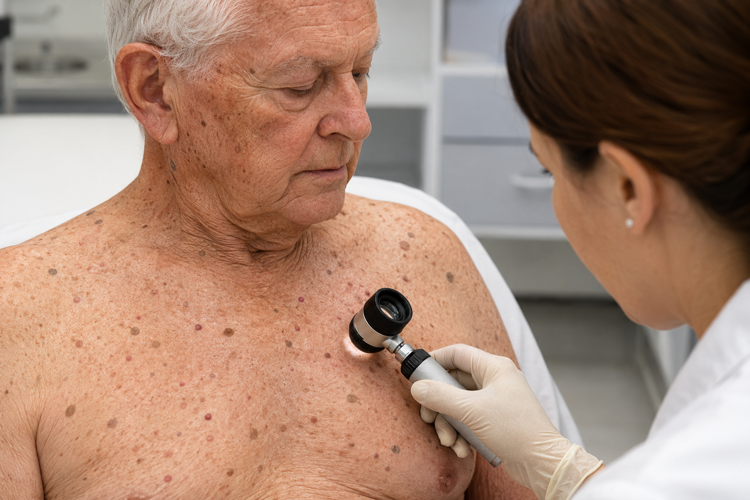
“Are These Dark Spots and Growths on My Skin Something to Worry About?”
A 78-year-old patient underwent a routine skin examination at Village Dermatology in Katy and Houston, Texas, revealing common benign skin growths including seborrheic keratoses, cherry angiomas, skin tags, and lentigines. Learn when these lesions should be evaluated and how dermatologists safely treat cosmetic concerns.
By: Dr. Caroline Vaughn
A Real Patient Case from Village Dermatology in Katy & Houston, Texas
As we age, it’s very common to notice new spots, bumps, and growths on the skin. Many patients worry these could be dangerous, but most are actually benign (non-cancerous) and part of normal skin aging.
This case highlights a 78-year-old male who presented for a routine upper body skin exam and was found to have several common, harmless skin growths.
Patient Case Overview
The patient came in for a full upper body skin check, with:
Asymptomatic lesions on the chest and neck
No pain, itching, or bleeding
On examination, several benign findings were identified:
Seborrheic keratoses (waxy, stuck-on growths)
Cherry angiomas (small red vascular spots)
Common Benign Skin Growths Explained
Seborrheic Keratoses
Waxy, “stuck-on” appearing lesions
Range from light tan to dark brown
Very common with aging
These are completely harmless and do not require treatment unless for cosmetic reasons.
Cherry Angiomas
Small, bright red or purple bumps
Caused by clusters of blood vessels
Increase in number over time
Also benign and do not require removal unless desired.
Skin Tags (Acrochordons)
Soft, flesh-colored growths
Common around the neck, armpits, and skin folds
Can become irritated from friction
In this case, multiple skin tags were safely removed with liquid nitrogen.
Lentigines (Sun Spots)
Flat, brown spots on sun-exposed areas
Caused by cumulative sun exposure over time
While harmless, they are a sign of sun damage, making sun protection essential.
Cosmetic Treatments Performed
Although these lesions are benign, some were treated for cosmetic reasons:
Liquid nitrogen (cryotherapy) used to remove:
1 seborrheic keratosis
10 skin tags
Patients were counseled on expected effects such as:
Temporary crusting or scabbing
Possible pigment changes
Low risk of scarring
The Importance of Sun Protection
Given the presence of sun-related skin changes, the patient was advised to:
Reapply every 2 hours when outdoors
Wear sun-protective clothing
Use SPF lip balm
Sunscreen helps prevent:
New sun spots
Skin aging
Skin cancer risk
When Should You See a Dermatologist?
Even though many skin growths are benign, it’s important to seek evaluation if you notice:
Rapid changes in size, shape, or color
Bleeding or non-healing lesions
New or unusual growths
Routine skin exams are especially important as we age.
Skin Cancer Screening & Dermatology Care in Katy & Houston, TX
Village Dermatology provides comprehensive skin exams and cosmetic treatments for patients across Katy and Houston, Texas, helping identify:
Benign vs. concerning lesions
Early signs of skin cancer
Age-related skin changes
Our goal is to keep your skin healthy, protected, and monitored over time.

“Why Does My Child Keep Getting These Itchy Bumps on Her Legs?”
A 4-year-old patient in Katy, TX presented with persistent itchy bumps diagnosed as molluscum contagiosum. Learn how dermatologists safely treat this common childhood condition.
By: Dr. Ashley Bladree
A Real Patient Case from Village Dermatology in Katy & Houston, Texas
Skin conditions in children can be stressful for parents—especially when bumps appear, spread, and don’t go away on their own. One of the most common causes of this in young children is molluscum contagiosum, a highly treatable viral skin condition.
This case highlights a 4-year-old female with persistent, itchy bumps on the back of her legs that required in-office treatment.
Patient Case Overview
The patient presented with:
Multiple itchy, irritated bumps on the back of both thighs
Symptoms present for several weeks
Increasing concern due to persistence and irritation
On exam:
Pink, shiny bumps with a central indentation (umbilication)
Classic appearance of molluscum contagiosum
Associated surrounding irritation (molluscum dermatitis)
What Is Molluscum Contagiosum?
Molluscum contagiosum is a common viral skin infection in children.
Key features:
Small, round, pink or flesh-colored bumps
Often have a central “dell” or indentation
Can be itchy or irritated
How it spreads:
Direct skin-to-skin contact
Shared items (towels, clothing)
Water exposure (pools, baths)
It is benign and very common, especially in children.
Why Do the Bumps Keep Spreading?
Many parents notice that the bumps increase over time.
This happens because:
The virus spreads easily through touch and scratching
Children may unknowingly autoinoculate (spread to other areas)
The immune system takes time to clear the virus
What Is Molluscum Dermatitis?
In this case, the patient also developed molluscum dermatitis, which is:
Red, irritated skin surrounding the bumps
Caused by the body’s immune response to the virus
This can make the condition appear worse—but it often signals that the body is starting to fight the infection.
Treatment Performed
Cantharidin (Blistering Therapy)
Applied to 8 lesions in the office
Causes controlled blistering to remove the bumps
Safe and commonly used in pediatric dermatology
Important Aftercare:
Wash off medication after 4 hours
Mild blistering or scabbing is expected
Avoid scratching to prevent spread
Supporting Treatment for Dermatitis
Triamcinolone cream for inflamed, itchy areas
Regular use of moisturizers (emollients) 2–3 times daily
When Should You Follow Up?
You should return if:
Bumps continue to spread rapidly
Lesions do not improve
Signs of infection appear (yellow crusting, pain)
This patient was scheduled for a 4-week follow-up to monitor progress.
Pediatric Dermatology Care in Katy & Houston, TX
Village Dermatology provides gentle, effective care for children with skin conditions like molluscum contagiosum across Katy and Houston, Texas. Our team focuses on:
Child-friendly treatment approaches
Minimizing discomfort and anxiety
Fast, effective clearance of lesions
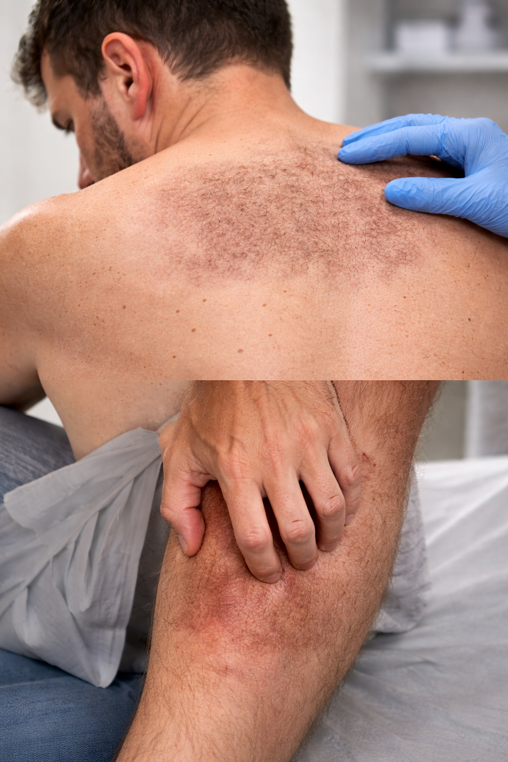
“Why Do I Still Have Dark Spots and Itchy Skin After My Rash Went Away?”
A 35-year-old patient in Katy, TX developed persistent dark spots and itching after a rash. Learn how dermatologists treat post-inflammatory hyperpigmentation and macular amyloidosis.
By: Dr. Ashley Baldree
A Real Patient Case from Village Dermatology in Katy & Houston, Texas
Many patients are surprised when a rash improves—but leaves behind dark patches or persistent itching. At Village Dermatology, we often see cases where the initial rash resolves, but secondary skin conditions like post-inflammatory hyperpigmentation (PIH) or macular amyloidosis remain.
This case highlights a 35-year-old male who developed lingering discoloration and itching after a rash triggered by insect bites.
Patient Case Overview
The patient presented with:
Red, itchy rash on the legs and trunk for 1 month
Partial improvement with topical steroids
Persistent dark patches and skin texture changes after the rash improved
On examination:
Hyperpigmented patches on the trunk
Mottled, rippled pigmentation on the upper back
Improvement of active rash, but residual skin changes
What Is Post-Inflammatory Hyperpigmentation (PIH)?
PIH is a common condition where the skin becomes darker after inflammation or injury.
Causes include:
Rashes
Insect bites
Scratching or irritation
Skin conditions like eczema or dermatitis
Key facts:
Discoloration can last months to years
More noticeable in patients with darker skin tones
Sun exposure can make it worse
In this case, PIH developed after the patient’s rash resolved.
What Is Macular Amyloidosis?
Macular amyloidosis is a lesser-known skin condition involving protein (amyloid) deposits in the skin.
Characteristics:
Brown, rippled or “reticulated” patches
Often located on the upper back
Associated with chronic friction or scratching
The patient’s history of itching and scratching likely contributed to this condition.
Why Is My Skin Still Itchy or Discolored?
Even after a rash improves:
The skin may remain inflamed beneath the surface
Scratching perpetuates the itch-scratch cycle
Pigment changes occur as part of the healing process
Breaking this cycle is key to recovery.
Treatment Plan and Recommendations
For Hyperpigmentation (PIH)
Sun protection (SPF 30+) daily
Minimize sun exposure
Consider laser therapy if persistent
For Macular Amyloidosis
Start AmLactin lotion to improve skin texture
Reduce friction and avoid scratching
Continue topical treatments as needed
For Residual Rash on Legs
Clobetasol cream twice daily for up to 2 weeks
Then use only as needed for flares
General Skin Care
Use gentle moisturizers (emollients)
Maintain hydration of the skin barrier
The Importance of Breaking the Itch-Scratch Cycle
This is one of the most important parts of treatment.
Scratching leads to:
More inflammation
Worsening pigmentation
Thickened or damaged skin
Stopping this cycle significantly improves outcomes.
When Should You Follow Up?
You should return if:
Pigmentation worsens or spreads
Itching persists despite treatment
New lesions develop
In this case, follow-up was scheduled in 4–6 weeks to monitor improvement.
Expert Skin Care in Katy & Houston, TX
Village Dermatology helps patients across Katy and Houston, Texas manage complex skin conditions like:
Post-inflammatory hyperpigmentation
Chronic itching disorders
Macular amyloidosis
Persistent rashes
Our approach focuses on accurate diagnosis, symptom control, and long-term skin health.
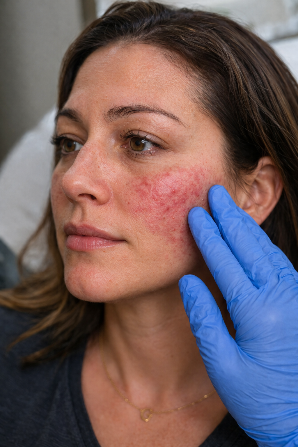
“Why Do I Still Get Rosacea Breakouts and Dark Spots Even When I’m Using My Cream?”
A 40-year-old patient in Katy, TX experienced persistent rosacea flares and dark spots despite treatment. Learn how dermatologists manage rosacea and prevent hyperpigmentation.
By: Dr. Caroline Vaughn
A Real Patient Case from Village Dermatology in Katy & Houston, Texas
Rosacea is one of the most common—and frustrating—chronic skin conditions we treat. Many patients feel discouraged when they are already using prescription creams but still experience flare-ups and lingering dark spots.
This case highlights a 40-year-old female dealing with persistent rosacea flares and post-inflammatory hyperpigmentation (PIH) despite appropriate topical therapy.
Patient Case Overview
The patient returned for follow-up after starting a triple rosacea cream (azelaic acid, metronidazole, and ivermectin).
She reported:
Improvement overall with her prescription cream
Continued intermittent inflammatory bumps
Concern about dark spots (PIH) after flares
On exam:
Mild inflammatory papules on the cheeks
Classic malar distribution of rosacea
Understanding Rosacea
Rosacea is a chronic inflammatory skin condition that primarily affects the central face.
Common symptoms:
Persistent redness
Acne-like bumps
Flushing and sensitivity
Visible blood vessels
Rosacea tends to flare and calm repeatedly, rather than fully resolve.
Why Am I Still Breaking Out?
This is one of the most common patient questions.
Even with good topical therapy:
Rosacea is chronic, not curable
Topicals may not fully control deeper inflammation
Triggers like heat, stress, alcohol, and spicy foods can still cause flares
In this case, the patient’s cream helped—but wasn’t enough to fully suppress inflammation.
What About the Dark Spots (PIH)?
Post-inflammatory hyperpigmentation (PIH) can occur after rosacea flares.
Important points:
PIH develops after inflammation heals
Treating PIH aggressively can irritate skin and worsen rosacea
The best strategy is preventing flares first
That’s why we focused on better inflammation control before targeting pigmentation.
Updated Treatment Plan
To improve long-term control, we adjusted her regimen:
Oral Anti-Inflammatory Therapy
Helps reduce inflammation without acting as a traditional antibiotic
Sulfur-Based Cleanser
Helps reduce bacteria and inflammation
Particularly effective for rosacea-prone skin
Continue Triple Cream
Azelaic acid
Metronidazole
Ivermectin
Essential Skin Care Tips for Rosacea
We reinforced gentle, consistent skincare:
Choose gentle cleansers and moisturizers
Green-tinted moisturizers can help reduce visible redness
Avoid harsh exfoliants or irritating products
Common Rosacea Triggers to Avoid
Patients should monitor and minimize:
Sun exposure
Heat and hot showers
Alcohol
Spicy foods
Stress
When Should You Follow Up?
You should return if:
Flares continue despite treatment
Symptoms worsen
You develop deeper nodules or cysts
In this case, follow-up was scheduled in 4–5 months to assess improvement.
Expert Rosacea Care in Katy & Houston, TX
Village Dermatology specializes in managing chronic rosacea and sensitive skin conditions, helping patients across Katy and Houston, Texas achieve clearer, calmer skin with:
Customized combination therapies
Medical and cosmetic treatment options
Long-term skin health strategies
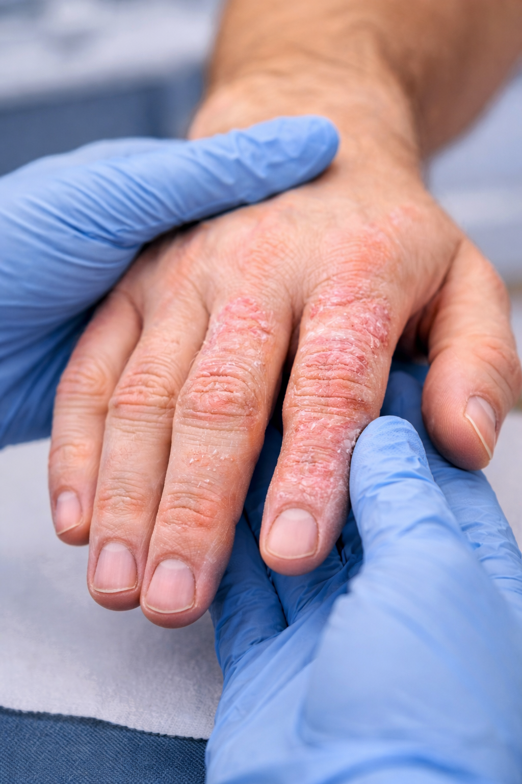
“Why Does My Hand Rash Keep Coming Back Even After Treatment?”
A 52-year-old patient in Katy, TX struggled with a recurring hand rash initially thought to be psoriasis. Learn how contact dermatitis is diagnosed and treated effectively.
By: Dr. Caroline Vaughn
A Real Patient Case from Village Dermatology in Katy & Houston, Texas
Chronic hand rashes can be frustrating—especially when they seem to improve, only to flare up again weeks later. At Village Dermatology, we frequently evaluate patients with persistent hand dermatitis, which is often mistaken for other skin conditions like psoriasis.
This case highlights a 52-year-old male with a recurring rash on his fingers that required careful diagnosis and targeted treatment.
Patient Case Overview
The patient presented with:
Itchy, moderate rash on the fingers
Involvement of the right index and middle fingers and left index finger
Symptoms recurring intermittently since 2020
Relevant History:
Previously diagnosed with dyshidrotic eczema
Later told it could be psoriasis, but treatment was inconsistent
Rash resolved temporarily, then returned after about 6 weeks
Recently prescribed clobetasol (high-potency topical steroid)
Final Diagnosis: Contact Dermatitis
After a detailed evaluation, the presentation was more consistent with contact dermatitis, rather than psoriasis.
Why psoriasis was less likely:
Limited to only a few fingers
No nail involvement
No joint pain (rules against psoriatic arthritis)
No widespread plaques elsewhere on the body
This pattern strongly suggests external irritation or allergic exposure as the root cause.
What Is Contact Dermatitis?
Contact dermatitis is a skin reaction caused by exposure to irritants or allergens.
Common triggers include:
Soaps and cleansers
Fragrances and skincare products
Metals (nickel)
Occupational exposures (chemicals, gloves, water)
Symptoms:
Red, itchy rash
Dry, cracked skin
Burning or irritation
Why Does It Keep Coming Back?
Many patients ask this exact question.
The answer: ongoing exposure to triggers
Even with treatment:
If the irritant isn’t removed, the rash will return
Some products labeled “moisturizing” can actually worsen irritation
Frequent handwashing can damage the skin barrier
In this case, certain skincare products were likely contributing to recurrence.
Treatment Plan and Recommendations
Topical Steroid Therapy
Continue clobetasol 0.05% as prescribed
Helps reduce inflammation and itching
Moisturizer Optimization
Switch to fragrance-free, hypoallergenic options:
Eucerin
Neutrogena Norwegian Formula
Continue O’Keeffe’s as needed
Discontinue potential irritants like certain scented products
Skin Care Routine
Use Dove sensitive skin soap
Avoid harsh soaps like Dial
Apply moisturizer after every hand wash
Apply steroid after shower and once more during the day
Overnight Repair
Apply Vaseline with cotton gloves overnight to restore skin barrier
When Is Patch Testing Needed?
If symptoms persist, patch testing may be recommended to identify specific allergens causing the reaction.
When Should You See a Dermatologist?
You should seek care if:
Rash lasts more than a few weeks
Symptoms keep recurring
Over-the-counter treatments are ineffective
Skin becomes cracked, painful, or infected
Expert Hand Dermatitis Treatment in Katy & Houston, TX
Village Dermatology provides expert care for chronic rashes and hand dermatitis, helping patients across Katy and Houston, Texas identify triggers and achieve long-term relief through personalized treatment plans.
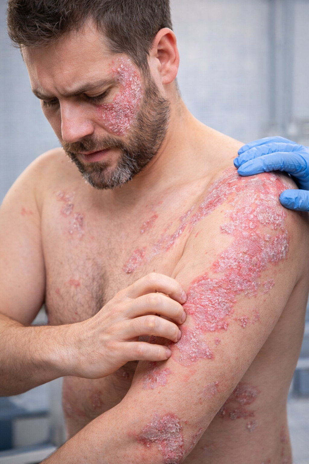
“Why Is My Psoriasis Getting Worse Even After Trying Biologic Treatment?”
A 35-year-old patient in Katy, TX experienced worsening psoriasis despite prior biologic therapy. Learn how advanced treatments like Skyrizi can help manage severe psoriasis.
By: Dr. Ashley Baldree
A Real Patient Case from Village Dermatology in Katy & Houston, Texas
Psoriasis can be frustrating—especially when it flares despite prior advanced treatments. At Village Dermatology, we often help patients navigate moderate-to-severe psoriasis when standard therapies are no longer effective.
This case highlights a 35-year-old male with widespread psoriasis who previously failed biologic therapy and required a more advanced treatment approach.
Patient Case Overview
The patient presented with:
Flaking, itchy, scaly plaques on the face, trunk, and arms
Symptoms present for several months with worsening severity
Prior treatment with Humira (adalimumab) without sustained control
On exam:
Psoriasiform plaques with micaceous scale
Body Surface Area (BSA): 40% involvement
Itch severity: 8/10
Importantly, the patient denied joint pain, indicating no current signs of psoriatic arthritis.
What Is Psoriasis?
Psoriasis is a chronic autoimmune skin condition that speeds up skin cell turnover, leading to:
Thick, scaly plaques
Red or inflamed skin
Itching and discomfort
Common triggers include:
Stress
Infections (like strep throat)
Certain medications
Alcohol use
Psoriasis often follows a pattern of flares and remissions, requiring long-term management.
Why Do Some Treatments Stop Working?
Many patients ask why their psoriasis worsens even after biologic therapy.
The reality is:
The immune system can adapt or become less responsive to certain biologics over time
Missed or delayed doses can lead to disease flare-ups
Each biologic targets a different immune pathway, so switching may be necessary
In this case, the patient had been off Humira for over a year, contributing to a significant flare.
Advanced Treatment: Skyrizi (Risankizumab)
Given the severity and prior treatment failure, we initiated Skyrizi, a newer biologic therapy.
Why Skyrizi?
Targets IL-23, a key driver of psoriasis inflammation
Effective for moderate-to-severe psoriasis
Convenient dosing:
Week 0
Week 4
Then every 12 weeks
Supporting Treatments
While starting biologic therapy, we also recommended:
Topical Steroid
Triamcinolone ointment for flare control
Medicated Shampoo
Ketoconazole shampoo to manage scalp involvement
Skin Care Measures
Daily moisturizers
Controlled sun exposure
Anti-dandruff shampoos (zinc, selenium, tar)
Safety and Monitoring
Because biologics affect the immune system, proper screening is essential.
Baseline Labs Ordered:
Tuberculosis screening (QuantiFERON-TB Gold)
Hepatitis B & C testing
HIV screening
Complete blood count and metabolic panel
Ongoing Monitoring:
Annual TB testing
Watch for signs of infection
Patients are carefully counseled on risks such as immunosuppression and infection.
When Should You See a Dermatologist?
You should seek expert care if:
Psoriasis covers large areas of the body
Symptoms interfere with daily life
Treatments are no longer effective
You experience frequent or severe flares
Early intervention can significantly improve outcomes and quality of life.
Expert Psoriasis Care in Katy & Houston, TX
Village Dermatology provides advanced, personalized care for psoriasis patients across Katy and Houston, Texas, including:
Biologic therapy management
Comprehensive lab monitoring
Individualized treatment plans
Long-term disease control strategies
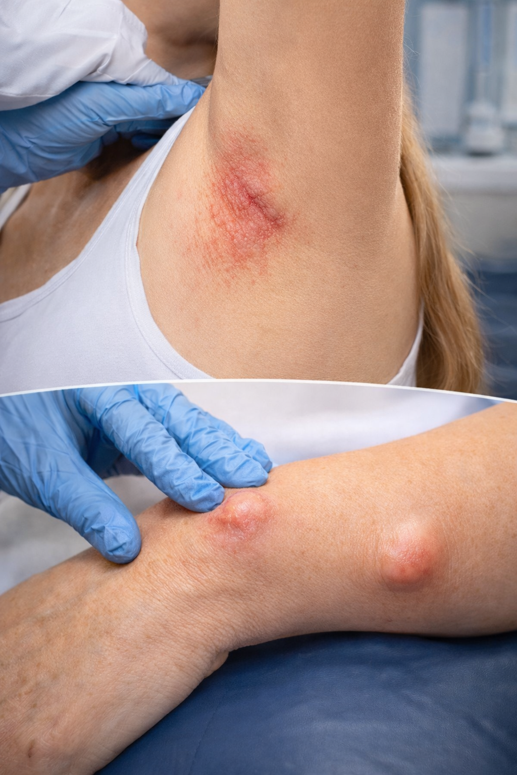
“Why Am I Getting Painful Red Skin Rashes and Bumps That Won’t Go Away?”
A 52-year-old patient in Katy, TX presented with persistent red rashes and painful cysts. Learn how intertrigo and epidermal cysts are diagnosed and treated by dermatologists.
By: Dr. Ashley Baldree
A Real Patient Case from Village Dermatology in Katy & Houston, Texas
Skin irritation in areas like the underarms, breast folds, or forearms can be frustrating—especially when symptoms persist for months. At Village Dermatology, we frequently evaluate patients with multiple skin concerns that may seem unrelated but are often connected.
This case highlights a 52-year-old female presenting with red skin lesions and a painful bump, ultimately diagnosed with intertrigo and an epidermal inclusion cyst.
Patient Case Overview
The patient came in with:
Red, inflamed skin lesions on the upper back, forearm, and underarm
A painful bump (cyst) on the forearm and breast
Symptoms present for several months without prior treatment
On examination:
The rash appeared consistent with intertrigo, a condition caused by friction and moisture
The bump was suspected to be an inflamed cyst vs. bug bite, later managed as a cyst
What Is Intertrigo?
Intertrigo is a common inflammatory skin condition that occurs in areas where skin rubs together, especially in warm, moist environments.
Common locations include:
Underarms
Under the breasts
Groin or abdominal folds
Symptoms:
Red, irritated patches
Burning or itching
Possible secondary fungal or bacterial infection
In this case, the patient’s underarm rash was consistent withintertrigo complicated by inflammation and possible fungal overgrowth.
What Is an Epidermal Inclusion Cyst?
An epidermal inclusion cyst is a benign, slow-growing lump beneath the skin filled with keratin.
Key features:
Round, firm bump under the skin
Can become painful, red, or infected
May require excision for complete removal
The patient’s cyst measured approximately 1 cm and caused discomfort, prompting treatment and planned removal.
Treatment Plan and Approach
At Village Dermatology, we take a comprehensive and targeted approach to treat multiple skin concerns simultaneously.
Intertrigo Treatment
Ketoconazole cream (antifungal) applied twice daily
Continued for 1 week after clearing to prevent recurrence
Recommended:
Barrier creams (zinc oxide/petrolatum)
Moisture control and friction reduction
Cyst Management (Forearm)
Vinegar soaks (1:1 vinegar + water) three times daily
Topical mupirocin antibiotic ointment applied after each soak
Cyst on Breast
Observation with plan for surgical excision if persistent
Why Do These Skin Conditions Occur Together?
Many patients are surprised to learn that:
Moisture, friction, and bacteria/fungus often work together to worsen skin conditions.
Intertrigo creates a compromised skin barrier
This environment allows microbial overgrowth
Cysts can become inflamed or infected in similar conditions
When Should You See a Dermatologist?
You should seek evaluation if:
Rashes persist for weeks or months
Skin becomes painful, swollen, or draining
A lump continues to grow or becomes tender
Early treatment can prevent complications and speed healing.
Expert Dermatology Care in Katy & Houston, TX
At Village Dermatology, we specialize in diagnosing and treating complex skin conditions, including rashes, infections, and cysts. Patients across Katy and Houston, Texas trust us for:
Accurate diagnosis with dermatoscopy
Personalized treatment plans
Medical and surgical dermatology expertise
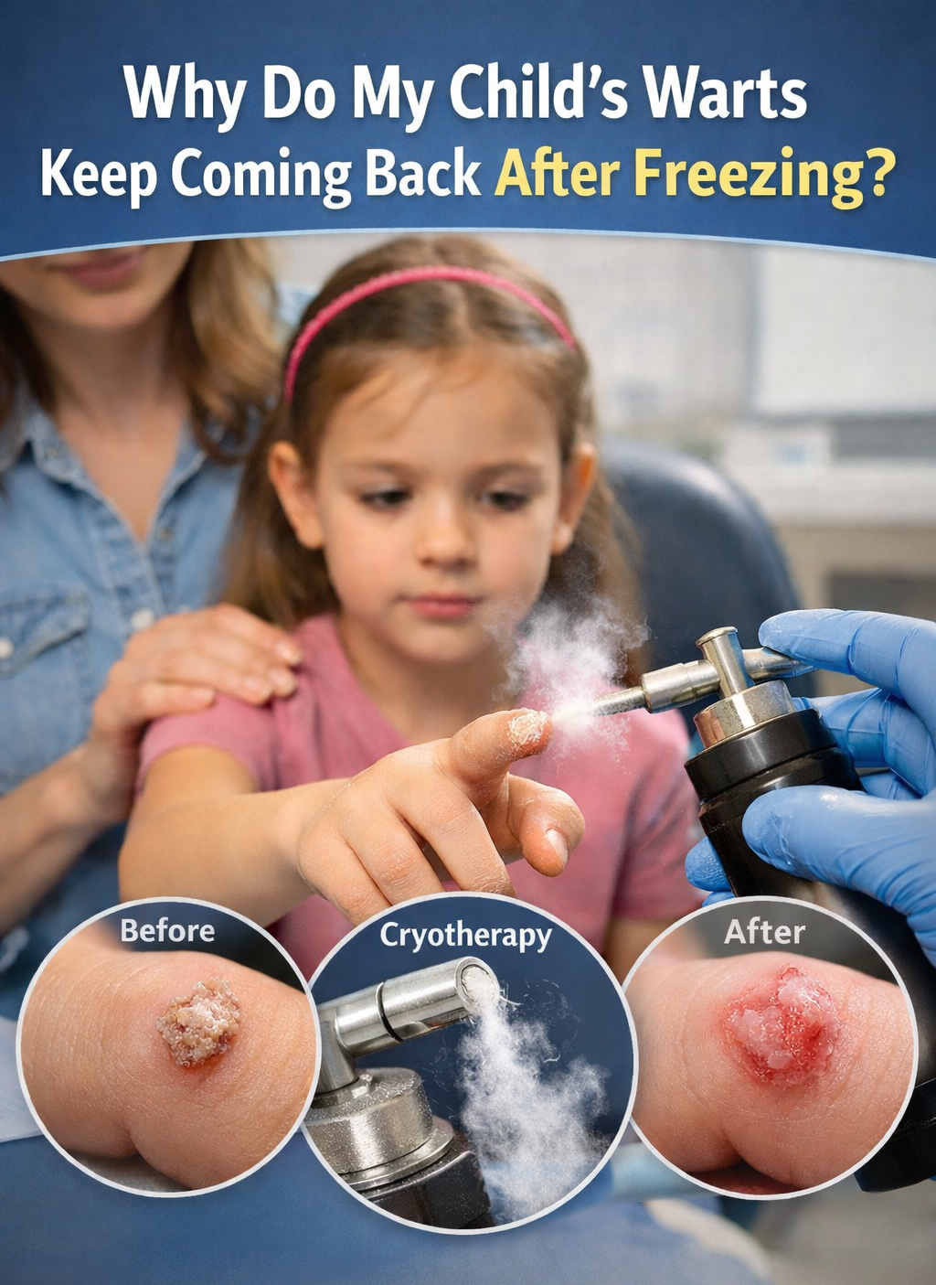
“Why Do My Child’s Warts Keep Coming Back Even After Freezing Them?”
A 7-year-old patient in Katy, TX presented with persistent warts despite cryotherapy. Learn why warts recur and how dermatologists in Houston effectively treat verruca vulgaris.
A Real Patient Case from Village Dermatology (Katy & Houston, Texas)
At Village Dermatology, we frequently see concerned parents asking why their child’s warts persist despite treatment. This recent case highlights a common—but treatable—skin condition known as verruca vulgaris (common warts).
Patient Case Overview
A 7-year-old female presented for follow-up evaluation of warts on her right hand, specifically on the ring finger and middle fingertip. She had previously undergone liquid nitrogen (cryotherapy) treatment about one month prior, with only mild improvement noted.
On examination, the lesions remained present and showed signs of:
Persistent growth
Mild inflammation
Thickened skin involvement near the nail
As part of treatment, careful trimming was performed to improve medication penetration, followed by another session of cryotherapy using liquid nitrogen.
What Are Verruca Vulgaris (Common Warts)?
Common warts are benign skin growths caused by the human papillomavirus (HPV). They often appear as:
Rough, cauliflower-like bumps
Skin-colored or slightly darker lesions
Found frequently on hands and fingers in children
They are contagious and can spread through:
Direct skin contact
Picking or scratching
Shared surfaces (e.g., towels, toys)
Why Didn’t the First Freezing Treatment Work Completely?
This is a very common concern. The truth is:
Warts often require multiple treatments.
Cryotherapy works by freezing the wart tissue, but:
Warts can extend deeper beneath the skin
The virus may persist even after visible improvement
Children’s immune systems respond at different speeds
In this case, the patient showed partial improvement, which is expected after just one session.
Treatment Approach at Village Dermatology
For this patient, we performed:
Cryotherapy (Liquid Nitrogen)
2 lesions treated
2 freeze–thaw cycles applied
Targeted destruction of wart tissue
Nail Trimming for Better Penetration
Helps treatment reach deeper viral tissue
Especially important for warts near or under nails
Education & Counseling
Families were advised that:
Multiple sessions are often needed
Warts may temporarily blister or scab after treatment
Recurrence is possible but manageable
Other Treatment Options for Warts
Depending on the case, we may also recommend:
Salicylic acid treatments (topical therapy)
Aldara (imiquimod cream)
Combination therapies for resistant warts
When Should You Follow Up?
You should return or contact your dermatologist if:
Warts are spreading quickly
They become painful or inflamed
There is no improvement after multiple treatments
In this case, the patient was scheduled for a 1-month follow-up to reassess response.
Why Choose Village Dermatology in Katy & Houston, TX?
At Village Dermatology, we specialize in treating pediatric and adult skin conditions with evidence-based, personalized care. Families across Katy and Houston, Texas trust us for:
Gentle pediatric dermatology care
Advanced wart removal techniques
Clear guidance for parents and patients
Compassionate, expert providers
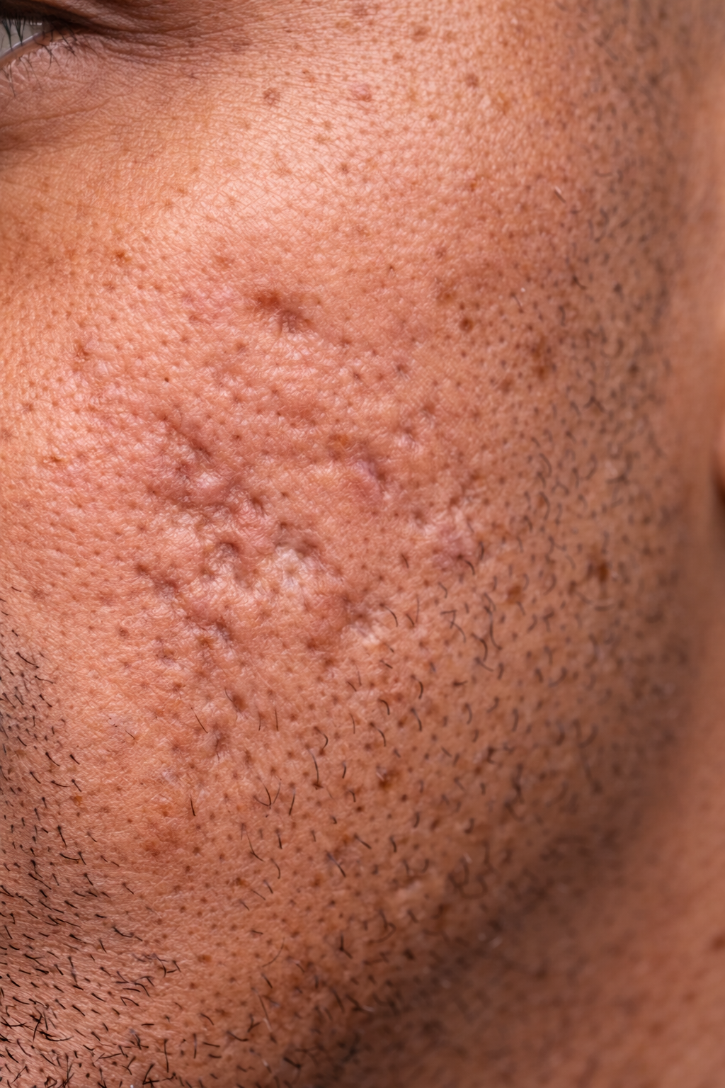
“Why Do I Still Have Acne Scars Years Later—and Can They Actually Be Fixed?”
A 31-year-old patient visited Village Dermatology in Katy, Texas for long-standing acne scars. Learn how dermatologists treat acne scarring with CO2 laser, TCA CROSS, and PRP.
At Village Dermatology in Katy, Texas and Houston, Texas, one of the most common concerns we hear from patients is about acne scars that never seem to go away.
A 31-year-old male patient recently came to our clinic frustrated with long-standing acne scars on both cheeks. He had struggled with acne in the past and was left with noticeable scarring that had persisted for years.
During the visit, he asked a very relatable question:
“Why do I still have acne scars years later—and is there anything that can actually fix them?”
Understanding Acne Scarring
On examination, the patient had moderate to severe rolling acne scars on both cheeks.
These scars form when:
Deep inflammation damages the skin
Collagen is lost during healing
The skin surface becomes uneven or depressed
Rolling scars typically appear as:
Wave-like depressions in the skin
Uneven texture
Shadowing on the cheeks
Unlike active acne, scars are permanent structural changes in the skin, which is why they require specialized treatments.
Why Acne Scars Don’t Go Away on Their Own
Many patients expect scars to fade completely over time, but this is not always the case.
While some improvement can occur in the first 1–2 years, deeper scars often persist because:
The skin has lost underlying support (collagen)
The healing process was incomplete
Repeated inflammation worsened damage
This is why professional dermatologic treatments are often needed.
Previous Treatment: CO2 Laser
This patient had previously undergone two CO2 laser treatments, with the last session over a year ago.
He reported:
Significant improvement after treatment
But still had residual scarring
This is very common—most patients require multiple treatment modalities for optimal results.
Treatment Options for Acne Scars
We discussed several advanced treatment options to further improve his skin.
TCA CROSS
A targeted chemical reconstruction technique used for:
Deep acne scars
Ice-pick and rolling scars
It works by:
Stimulating collagen production
Gradually improving scar depth
PRP (Platelet-Rich Plasma)
PRP uses the patient’s own blood to:
Promote healing
Boost collagen production
Enhance results when combined with other treatments
CO2 Laser Resurfacing
A highly effective treatment that:
Resurfaces the skin
Stimulates new collagen
Improves overall texture
Often requires multiple sessions for best results.
Why Combination Treatment Works Best
For moderate to severe acne scarring, a combination approach is often recommended.
In this case, the patient was quoted a treatment plan including:
TCA CROSS
PRP
CO2 laser resurfacing
These treatments work together to:
Target different scar depths
Improve overall skin texture
Deliver more noticeable results
What About Topical Treatments?
We also discussed topical options such as tretinoin, which can help improve skin texture over time.
However, the patient reported sensitive skin and irritation with topicals, so we recommended:
OTC Adapalene (Differin Gel)
A milder retinoid
Helps with skin turnover
May improve texture gradually
Patients should discontinue use if irritation occurs.
Realistic Expectations for Acne Scar Treatment
It’s important for patients to understand:
Acne scars cannot be completely erased
Treatments aim to significantly improve appearance
Results take time and multiple sessions
Gradual improvement is expected over months
Consistency and patience are key.
When to See a Dermatologist
You should consider treatment if:
Acne scars affect your confidence
Skin texture is uneven
Over-the-counter products are not helping
You want more advanced cosmetic improvement
Acne Scar Treatment in Katy and Houston, Texas
At Village Dermatology, we offer advanced treatments for acne scarring including:
CO2 laser resurfacing
TCA CROSS
PRP therapy
Customized treatment plans
If you are struggling with persistent acne scars, our dermatology team can help you explore effective options for smoother, healthier skin.
“My Moles Haven’t Changed… So Why Do I Still Need to Get Them Checked?”
A 52-year-old patient followed up at Village Dermatology in Katy, Texas for stable moles. Learn why dermatologists still recommend routine skin checks even when moles haven’t changed.
By: Dr. Ashley Baldree
At Village Dermatology in Katy, Texas and Houston, Texas, one of the most common questions patients ask during follow-up visits is:
“If my moles look the same, do I really need to keep checking them?”
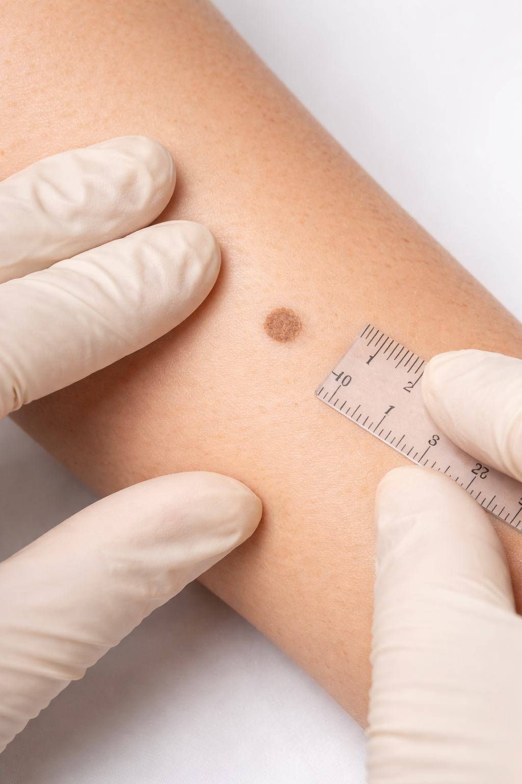
A 52-year-old female patient recently came in for a follow-up evaluation of benign moles (nevi) located on her body, including her left forearm and upper back. These moles had previously been measured and documented during her last visit.
Why Follow-Up Visits for Moles Matter
During this visit, a detailed skin examination was performed using a dermatoscope, allowing for close evaluation of the patient’s moles.
The previously monitored lesions measured:
4 mm on the left forearm
4.5 mm on the upper back
Importantly, both lesions remained:
Symmetrical
Evenly colored
Stable in size
These findings are consistent with benign nevi, meaning the moles are non-cancerous and do not require treatment.
However, even stable moles should continue to be monitored over time.
Why You Still Need to Monitor “Normal” Moles
Even when moles appear unchanged, dermatologists recommend continued surveillance because:
Skin changes can happen gradually and subtly
New moles or lesions may develop
Early skin cancer can mimic benign moles
A baseline comparison helps detect future changes
Regular monitoring ensures that any concerning changes are caught early, when treatment is most effective.
What Are Benign Nevi?
Benign nevi are extremely common and typically appear as:
Small brown or tan spots
Round or oval in shape
Evenly pigmented
Smooth borders
Most adults have multiple moles, and the majority remain harmless throughout life.
Other Common Skin Findings
During the exam, the patient also had additional benign skin conditions.
Lentigines (Sun Spots)
Lentigines are light brown spots caused by cumulative sun exposure.
They are commonly found on:
Arms
Face
Chest
Back
While harmless, they can be improved cosmetically with:
Retinoids
Chemical peels
Daily sun protection is key to preventing new spots.
Sebaceous Hyperplasia
The patient also had sebaceous hyperplasia on the cheek.
These appear as:
Small yellow or flesh-colored bumps
Enlarged oil glands
Dome-shaped papules
They are completely benign and do not require treatment, but can be removed if desired using:
Laser therapy
Electrodessication
How to Check Your Moles at Home
Patients were advised to perform monthly self-skin exams.
Use the ABCDE rule when evaluating moles:
A – Asymmetry
B – Border irregularity
C – Color variation
D – Diameter (larger than 6 mm)
E – Evolving (changing over time)
If any mole changes in size, shape, color, or begins to itch or bleed, it should be evaluated promptly.
The Role of Sunscreen in Skin Health
Patients were strongly encouraged to use:
Sun-protective clothing when outdoors
Sunscreen helps:
Prevent new moles and sun spots
Reduce skin cancer risk
Protect against premature aging
When Should You See a Dermatologist?
You should schedule a skin exam if you notice:
New moles
Changes in existing moles
Spots that itch, bleed, or grow
Any lesion that looks different from others (“ugly duckling sign”)
Even without changes, annual skin checks are recommended.
Dermatology Care in Katy and Houston, Texas
At Village Dermatology, we specialize in:
Full-body skin exams
Mole monitoring and dermoscopy
Skin cancer screening and prevention
Treatment of benign skin lesions
If you have concerns about your moles or skin spots, our dermatology team is here to help.
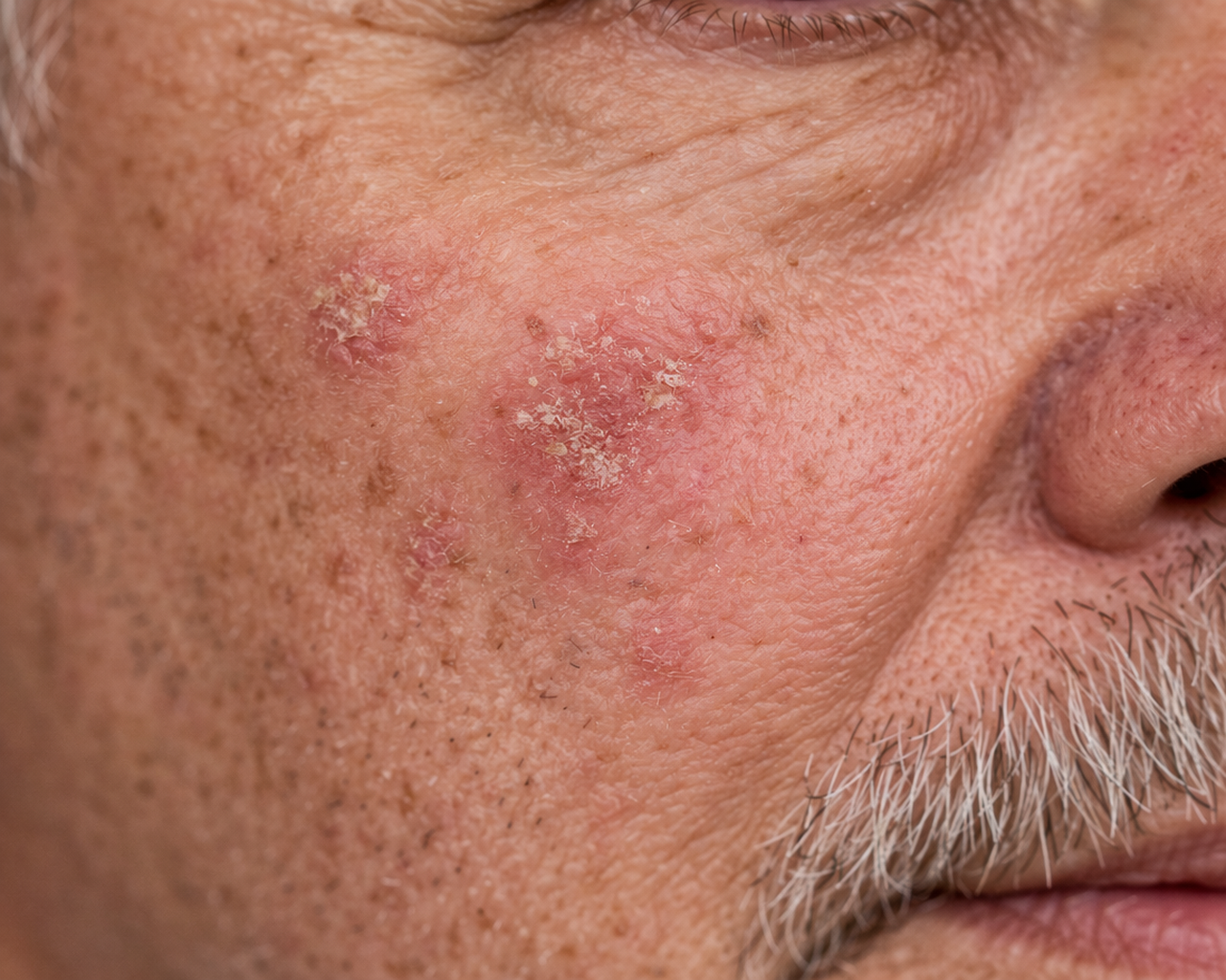
“Do I Still Need Treatment If My Pre-Cancer Spots Look Better After PDT?”
A 63-year-old patient followed up at Village Dermatology in Katy, Texas after PDT treatment for actinic keratoses. Learn whether additional treatment is needed and how to prevent recurrence.
By: Dr. Ashley Baldree
At Village Dermatology in Katy, Texas and Houston, Texas, many patients return after treatment for precancerous skin lesions wondering what comes next. A 63-year-old male patient recently came in for a follow-up visit after undergoing photodynamic therapy (PDT) for actinic keratoses on the face.
At his appointment, he asked a very common and important question:
“Do I still need treatment if my pre-cancer spots look better after PDT?”
Follow-Up After Photodynamic Therapy (PDT)
This patient had previously been treated with red light photodynamic therapy, a highly effective treatment for actinic keratoses (AKs).
During his follow-up visit:
There was significant improvement
Previously visible lesions had markedly reduced
The patient reported no complications from treatment
Because of this excellent response, no additional treatment was needed at this time, and the plan was to continue routine monitoring.
What Are Actinic Keratoses?
Actinic keratoses are precancerous skin lesions that develop due to long-term sun exposure.
They typically appear as:
Rough, scaly patches
Red or pink spots
Areas that may feel like sandpaper
AKs are important to treat because a small percentage can progress to:
Squamous Cell Carcinoma (SCC)
Early treatment and follow-up significantly reduce this risk.
Do You Need Treatment After PDT?
Even when lesions improve or disappear, ongoing monitoring is essential.
Why?
New AKs can develop over time
Sun damage is cumulative
Some lesions may recur
If no active lesions are present, dermatologists often recommend:
Observation
Routine skin exams
Sun protection
Treatment is only restarted if new lesions appear.
How to Prevent Actinic Keratoses from Returning
Patients were counseled on the importance of sun protection:
Daily Prevention Tips
Use broad-spectrum sunscreen SPF 30+ every day
Wear sun-protective clothing and hats
Avoid peak sun hours when possible
Reapply sunscreen every 2 hours outdoors
Consistent sun protection is the most effective way to prevent recurrence.
Additional Diagnosis: Rosacea
During the visit, the patient was also noted to have rosacea, a chronic inflammatory skin condition.
Symptoms included:
Redness
Acne-like bumps (papules and pustules)
Facial sensitivity
Treatment Plan for Rosacea
To help manage rosacea, the patient was started on:
Oral Doxycycline 20 mg
Taken twice daily with food
Helps reduce inflammation and breakouts
Topical Triple Cream
Applied nightly to the face
Helps control redness and lesions
Rosacea Triggers to Avoid
Patients were advised that rosacea can flare with:
Sun exposure
Heat
Spicy foods
Alcohol
Stress
Wind
Avoiding triggers can significantly reduce flare-ups.
When to Follow Up
The patient was scheduled to return in 3 months to reassess both:
Patients should return sooner if they notice:
New rough or scaly spots
Persistent redness or worsening bumps
Painful or non-healing lesions
Dermatology Care in Katy and Houston, Texas
At Village Dermatology, we specialize in:
Actinic keratosis treatment
Photodynamic therapy (PDT)
Rosacea management
Skin cancer prevention
Comprehensive skin exams
If you have sun-damaged skin, rough spots, or facial redness, our dermatology team can help you maintain healthy skin.
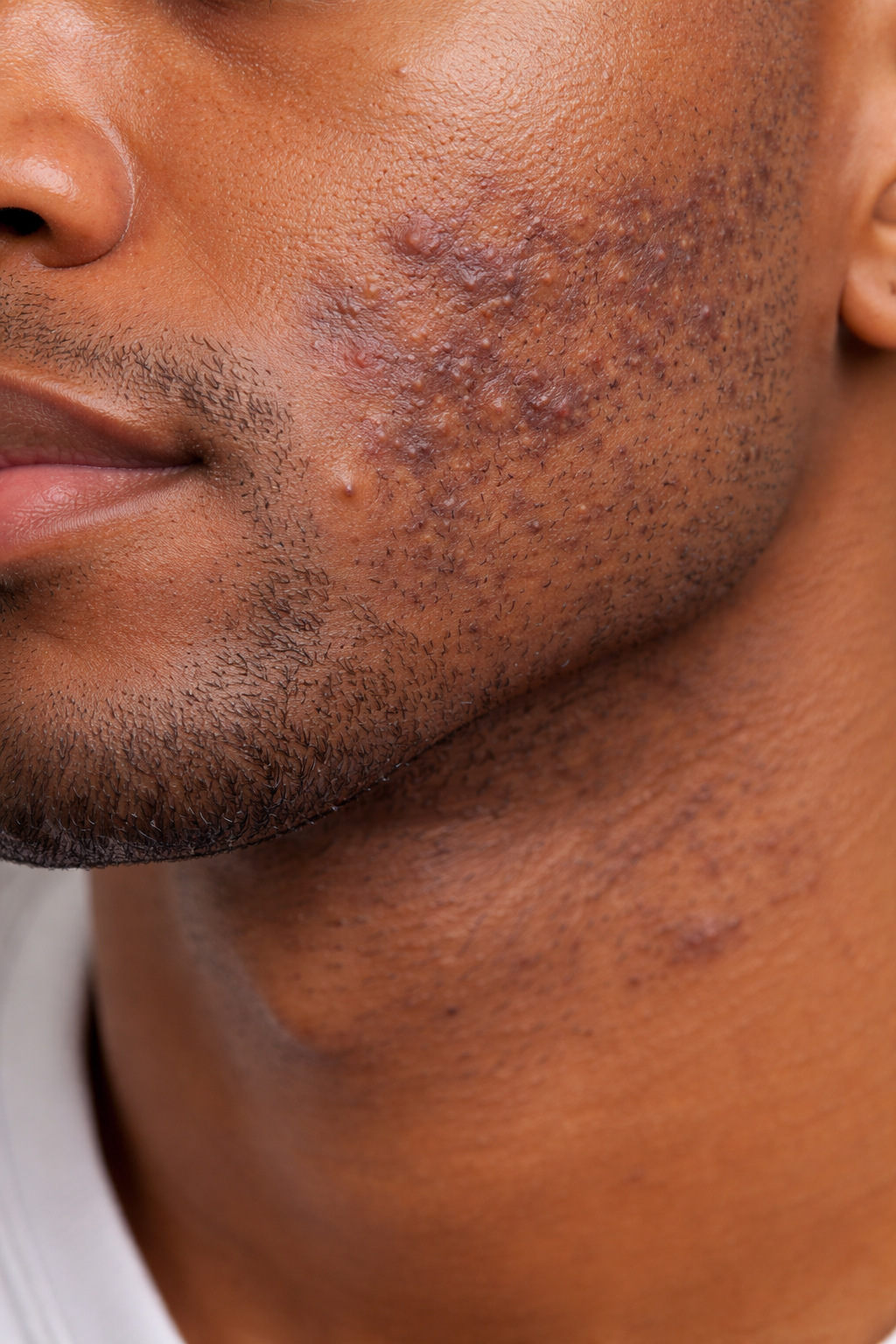
“How Do I Get Rid of Razor Bumps and Dark Spots on My Face?”
A 24-year-old patient visited Village Dermatology in Katy, Texas for dark spots and razor bumps. Learn how dermatologists treat pseudofolliculitis barbae and post-inflammatory hyperpigmentation.
At Village Dermatology in Katy, Texas and Houston, Texas, many young patients come in with concerns about dark spots and bumps on the face, especially after shaving.
A 24-year-old male patient recently visited our clinic with complaints of:
Brown discoloration on the cheeks
Recurrent bumps after shaving
Persistent skin changes that were not improving on their own
His main question during the visit was:
“How Do I Get Rid of Razor Bumps and Dark Spots on My Face?”
This is a very common concern, especially in patients with sensitive or curly facial hair.
Understanding Facial Hyperpigmentation
On examination, the patient had hyperpigmented patches on both cheeks, which are darker than the surrounding skin.
This condition is often referred to as post-inflammatory hyperpigmentation (PIH).
PIH occurs when the skin produces excess pigment after:
Irritation
Inflammation
Shaving-related trauma
These dark spots can take months to years to fade, especially without proper treatment and sun protection.
What Is Pseudofolliculitis Barbae (Razor Bumps)?
In addition to discoloration, the patient also had pseudofolliculitis barbae, commonly known as razor bumps.
This condition occurs when:
Hair curls back into the skin after shaving
The body reacts with inflammation
Small red or dark bumps form on the skin
It is most common in individuals with curly or coarse hair.
Symptoms may include:
Painful or itchy bumps
Dark spots after healing
Ongoing irritation with shaving
Why Razor Bumps Cause Dark Spots
Every time the skin becomes inflamed from ingrown hairs, it can leave behind post-inflammatory hyperpigmentation.
This creates a cycle:
Shaving causes irritation
Razor bumps develop
Skin heals with dark spots
New shaving leads to repeat irritation
Breaking this cycle is key to improving both bumps and discoloration.
Treatment Plan for Razor Bumps and Dark Spots
We developed a treatment plan to address both inflammation and pigmentation.
Morning Routine
Benzoyl Peroxide Wash (PanOxyl or CeraVe)
Helps reduce bacteria and inflammationClindamycin Gel
A topical antibiotic that treats inflamed bumpsSunscreen (SPF 30+)
Prevents dark spots from worsening
Evening Routine
Gentle Cleanser (La Roche-Posay)
Keeps skin clean without irritationTretinoin Cream
Helps by:Increasing skin turnover
Preventing clogged pores
Fading dark spots over time
Moisturizer (if needed)
Helps reduce dryness from treatment
Shaving Tips to Prevent Razor Bumps
Patients were counseled on proper shaving techniques to reduce irritation:
Shave with the grain, not against it
Avoid shaving too closely
Use clean, sharp razors
Consider electric clippers instead of razors
Avoid repeated passes over the same area
For long-term improvement, laser hair removal may be considered, as it can reduce hair growth and prevent ingrown hairs.
How Long Does It Take to See Improvement?
Patients should expect:
Improvement in bumps within a few weeks
Gradual fading of dark spots over several months
Continued improvement with consistent skincare and sun protection
If symptoms persist, additional treatments may be recommended.
Dermatology Care in Katy and Houston, Texas
At Village Dermatology, we specialize in treating:
Razor bumps (pseudofolliculitis barbae)
Hyperpigmentation
Acne and post-inflammatory skin changes
Chronic facial irritation
If you are experiencing persistent bumps or dark spots on your face, our dermatology team can create a personalized treatment plan to restore healthy skin.
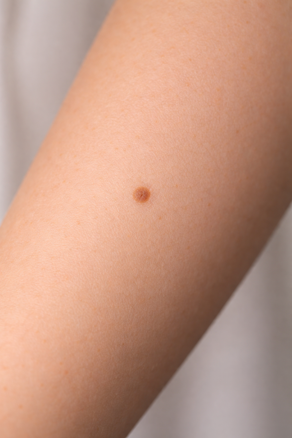
“Should I Be Worried If My Moles Haven’t Changed Since My Last Skin Check?”
Concerned about moles or sun spots? Village Dermatology in Katy and Houston, Texas offers expert mole checks, skin cancer screenings, and dermatologic evaluations for suspicious skin lesions.
At Village Dermatology in Katy, Texas and Houston, Texas, many patients return for follow-up visits to monitor their moles and other skin spots. One of the most common questions dermatologists hear during these visits is:
“If my moles look the same as last year, do I still need to keep checking them?”
A 52-year-old female patient recently came in for a follow-up visit after previously being evaluated for benign moles (nevi) located on her body, including her left forearm and upper back.
She had been seen several months earlier, and photographs were taken to monitor the lesions. At this visit, the goal was to ensure the moles remained stable and showed no signs of skin cancer.
Monitoring Moles Over Time
During the follow-up visit, a dermatologic examination was performed using a dermatoscope, a specialized magnifying tool dermatologists use to examine skin lesions in detail.
The patient’s moles were carefully evaluated and documented.
Two specific lesions that were previously monitored included:
Left distal dorsal forearm – 4 mm mole
Left medial upper back – 4.5 mm mole
Both lesions remained:
Regular in shape
Symmetrical
Evenly pigmented
These characteristics are consistent with benign nevi, meaning the moles are non-cancerous and stable.
Because the lesions had not changed, the recommended approach was continued observation.
What Are Benign Nevi?
Benign nevi are very common skin growths composed of clusters of pigment-producing cells.
They typically appear as:
Small brown or tan spots
Evenly colored macules or papules
Symmetrical lesions with smooth borders
Most adults have 10 to 40 moles on their body, and they are usually harmless.
However, dermatologists recommend monitoring moles because changes over time can signal early skin cancer, particularly melanoma.
Why Dermatologists Take Photographs of Moles
At Village Dermatology, clinical photos may be taken to help monitor moles over time.
This allows dermatologists to:
Compare lesions during future visits
Detect subtle changes early
Avoid unnecessary biopsies when lesions remain stable
This approach is especially helpful for patients with multiple moles.
Other Common Skin Findings
In addition to benign moles, this patient had other common and harmless skin conditions.
Lentigines (Sun Spots)
Lentigines are light tan or brown spots caused by sun exposure.
They often appear on sun-exposed areas such as:
Face
Arms
Chest
Back
Although they are harmless, some patients choose treatment for cosmetic reasons.
Possible treatments include:
Retinoid creams
Chemical peels
Laser treatments
Skin-brightening products
Daily broad-spectrum sunscreen SPF 30 or higher is essential to prevent new spots from forming.
Sebaceous Hyperplasia
Another finding during the visit was sebaceous hyperplasia, located on the patient’s cheek.
Sebaceous hyperplasia occurs when oil glands enlarge and appear as:
Small yellow or flesh-colored bumps
Soft dome-shaped papules
Often located on the face
These lesions are completely benign and do not require treatment.
However, if desired, they can be treated with:
Electrodesiccation
Laser therapy
Topical retinoids
How to Monitor Your Moles at Home
Patients were advised to perform monthly self-skin exams to look for any changes in their moles.
Dermatologists recommend following the ABCDE rule when checking moles:
A – Asymmetry
B – Border irregularity
C – Color changes
D – Diameter larger than 6 mm
E – Evolving (changing over time)
If a mole begins to:
Grow
Change color
Become irregular
Itch, bleed, or become painful
it should be evaluated by a dermatologist.
The Importance of Annual Skin Exams
Even when moles appear normal, annual skin exams with a dermatologist are recommended.
Routine skin checks help detect:
Early melanoma
Basal cell carcinoma
Squamous cell carcinoma
Other suspicious lesions
Early detection greatly improves treatment outcomes.
Skin Monitoring and Dermatology Care in Katy and Houston, Texas
At Village Dermatology, we provide comprehensive skin evaluations including:
Full-body skin exams
Mole monitoring
Skin cancer screenings
Dermatoscopic evaluation
Treatment of benign and cosmetic skin lesions
If you have moles you want checked or changes in your skin, our dermatology team can help.
Schedule a skin exam at Village Dermatology in Katy or Houston, Texas to ensure your skin remains healthy.
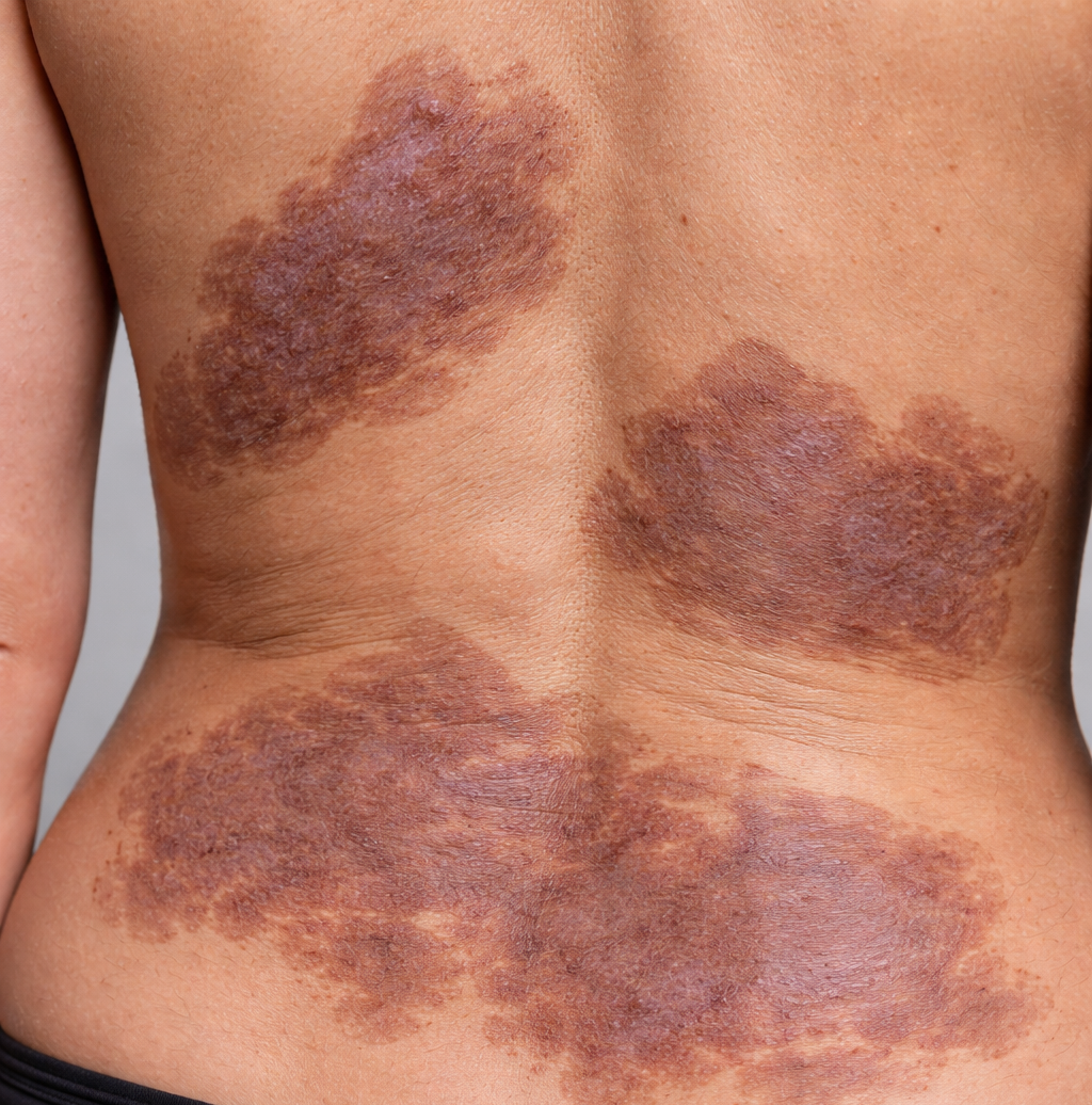
“Why Do I Have Dark, Thick Patches on My Skin That Won’t Go Away?”
Struggling with thickened or discolored skin? Village Dermatology in Katy and Houston, Texas offers expert treatment for morphea, chronic rashes, and skin pigmentation disorders.
At Village Dermatology in Katy, Texas and Houston, Texas, patients often come to us with long-standing skin conditions that have not improved with prior treatments. One such case involved a 43-year-old female patient who presented with a chronic rash affecting multiple areas of her body.
Her main concern during the visit was:
“Why do I have dark, thick patches on my skin that won’t go away?”
This is a common question among patients dealing with morphea, a rare but persistent skin condition.
Understanding Generalized Morphea
During the examination, the patient was found to have generalized morphea, a condition she had previously been diagnosed with and treated for using phototherapy and systemic medications.
On exam, she had:
Confluent, bound-down hyperpigmented plaques
Areas of skin thickening (sclerosis)
Lesions distributed across the back, trunk, and breasts
Morphea is a type of localized scleroderma, which causes:
Hardening and thickening of the skin
Changes in pigmentation
Long-lasting plaques that may persist for years
Unlike systemic scleroderma, morphea typically does not affect internal organs, which is reassuring for many patients.
Why Morphea Can Be Difficult to Treat
Morphea can be challenging because:
It is chronic and long-lasting
Response to treatment can vary significantly
Lesions may improve slowly over time
Some areas may remain permanently changed
Even with treatment, patients may experience periods of progression and stability.
Treatment Options for Morphea
This patient had previously tried:
Methotrexate
Other light-based treatments
Given her ongoing symptoms, we discussed restarting treatment with a structured approach.
Phototherapy (Light Treatment)
Phototherapy is one of the most effective treatments for morphea.
The plan included:
Starting in-office phototherapy sessions
Initiating the process for at-home phototherapy approval
Phototherapy helps by:
Reducing inflammation
Softening thickened skin
Slowing progression of plaques
This treatment is often used long-term to manage symptoms.
Topical Treatments
Patients may also benefit from:
Topical steroids to reduce inflammation
Calcipotriene to help regulate skin cell growth
These treatments can improve the appearance and texture of affected skin.
Photoaging and Skin Health
In addition to morphea, the patient also had photoaging (sun damage) on the face.
Photoaging can cause:
Uneven pigmentation
Fine lines and wrinkles
Thinning of the skin
To address this, we recommended:
Tretinoin Benefits
Tretinoin helps by:
Increasing skin cell turnover
Improving skin texture
Reducing pigmentation and fine lines
Patients should:
Apply a pea-sized amount at night
Start 2–3 times per week
Increase gradually as tolerated
Cosmetic Treatment Considerations
The patient also expressed interest in cosmetic treatments. Given her diagnosis of morphea, we discussed safe options.
Safe Options:
Treatments to Avoid:
Laser resurfacing
These more aggressive treatments may worsen skin changes in patients with morphea.
When to Follow Up
Because morphea can evolve over time, the patient was advised to return in 2–3 months to monitor progress and adjust treatment as needed.
Patients should also contact their dermatologist if:
Lesions spread
Skin becomes more firm or thickened
New areas of involvement appear
Dermatology Care in Katy and Houston, Texas
At Village Dermatology, we specialize in treating complex and chronic skin conditions such as:
Morphea (localized scleroderma)
Chronic rashes
Pigmentation disorders
Photoaging and sun damage
If you are experiencing persistent skin changes that do not improve, our dermatology team can provide expert evaluation and personalized treatment.