

Evaluating a Scalp Growth and Atopic Dermatitis in a 20-Year-Old Female — Village Dermatology Katy & Houston, Texas
A 20-year-old female presented to Village Dermatology in Katy and Houston, Texas with a scalp growth, eczema flare, and post-inflammatory hyperpigmentation. Learn how dermatologists assess suspicious lesions and manage chronic atopic dermatitis and PIH.
Young adults often present with a combination of dermatologic concerns involving both growths and inflammatory skin conditions. This case report highlights a 20-year-old female evaluated at Village Dermatology, serving Katy and Houston, Texas, for a scalp growth, eczema flares, and post-inflammatory hyperpigmentation (PIH).
Chief Complaints
Growth on the left central parietal scalp
Rash on the hands and left leg
The patient reported:
A moderate, asymptomatic growth on the scalp
Flaking rash on the hands and left lower leg
Concern about dark discoloration (PIH) left behind after rashes
She had been previously treating eczema with triamcinolone.
Clinical Examination
A focused examination evaluated the:
Scalp
Face
Hands
Left lower leg
The patient was well-appearing, alert, and in no distress.
Findings included:
1. Scalp Lesion
A solitary lesion on the left central parietal scalp concerning for:
Neoplasm of uncertain behavior
Nevus
Lipofibroma
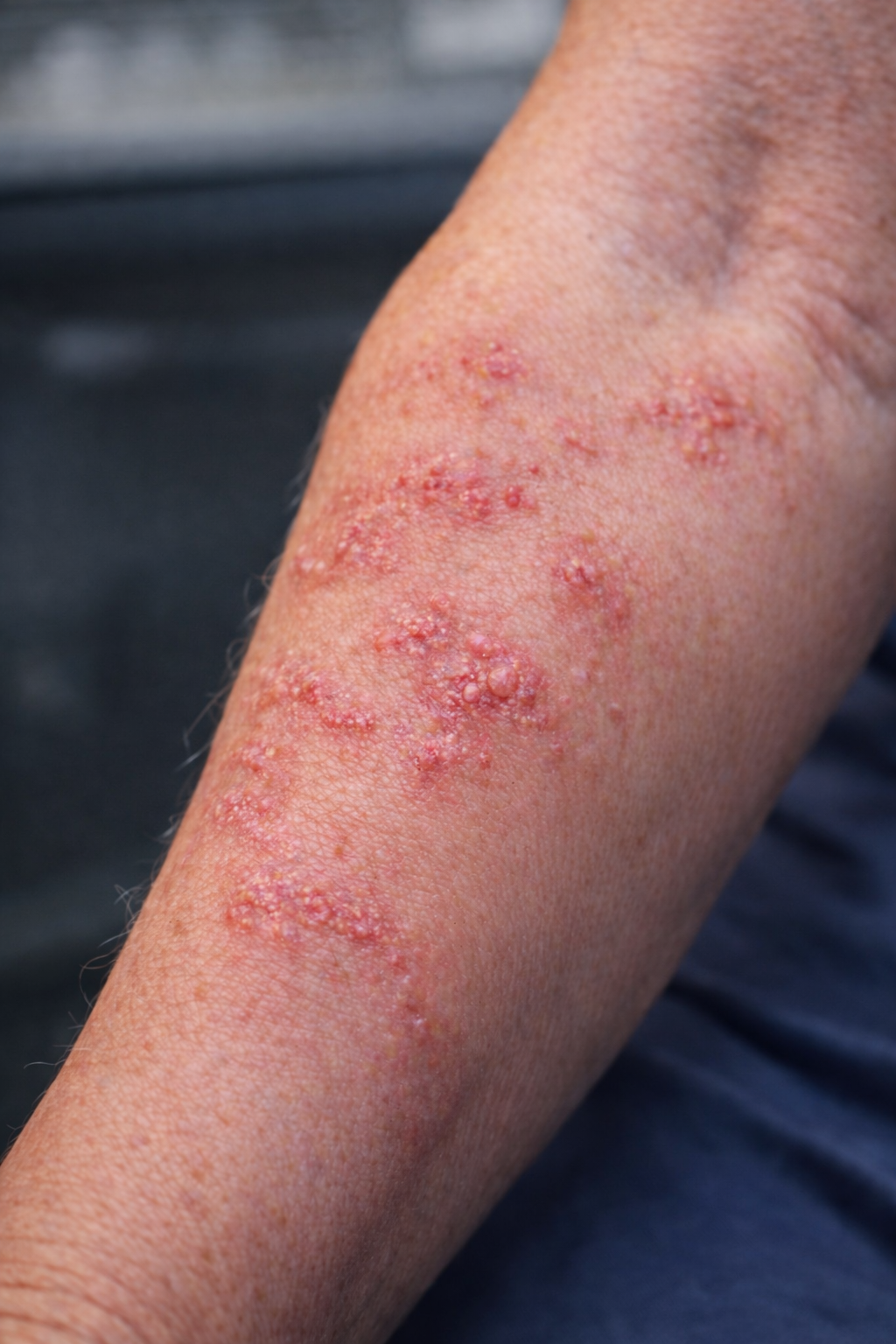
2. Active Atopic Dermatitis
Erythematous, eczematous patches on:
Left proximal pretibial region
Left ulnar dorsal hand
Right ulnar dorsal hand
3. Post-Inflammatory Hyperpigmentation
Ill-defined hyperpigmented patches in the same areas where eczema was present.
Diagnosis & Assessment
1. Neoplasm of Uncertain Behavior
Given the uncertain nature of the scalp growth, a biopsy was recommended.
Biopsy Procedure
Shave biopsy of the lesion
Local anesthesia with lidocaine + epinephrine
Dermablade used to obtain specimen for H&E
Hemostasis with Drysol
Petrolatum applied post-procedure
The patient will be notified of results within 2 weeks.
2. Atopic Dermatitis
The patient had persistent eczema despite 2 months of topical steroid use.
She was experiencing an active flare.
Treatment Plan
Fluocinonide 0.05% cream for the body
Fluocinonide 0.05% solution for the scalp
Daily moisturization with CeraVe cream
Discussed long-term options:
Continued topical therapy
Dupixent injections (biologic therapy)
Steroid counseling included:
Avoiding prolonged use
Avoiding high-potency steroids on face, groin, or skin folds
Possible side effects: atrophy, telangiectasias, hypopigmentation
3. Post-Inflammatory Hyperpigmentation (PIH)
PIH was present secondary to eczema flares.
Counseling Included:
PIH fades naturally but may take months to years
Strict sun protection recommended
Goal is first to control active eczema, then address pigmentation
Patient Counseling
Topics reviewed during the visit:
Skin Care for Eczema
Use lukewarm showers
Apply moisturizers immediately after bathing
Use unscented cleansers and detergents
Avoid excessive hand washing
Keep nails short to reduce scratching
When to Contact the Office
Worsening rash
Signs of infection (yellow crusts or cold sores)
Darkening or spreading hyperpigmentation
Follow-Up
The patient will return in 1 month for evaluation of:
Biopsy results
Eczema response to treatment
PIH improvement
Why Early Evaluation Matters
Young adults often overlook concerning skin growths or chronic rashes.
At Village Dermatology, serving Katy and Houston, we provide:
Expert evaluation of suspicious lesions
Personalized eczema treatment plans
Guidance on pigmentary disorders such as PIH
This case highlights the importance of comprehensive dermatologic care in patients with overlapping concerns.

Managing Longstanding Melasma in a 50-Year-Old Female — Village Dermatology Katy & Houston, Texas
Learn how Village Dermatology in Katy and Houston, Texas evaluated and treated a 50-year-old woman with longstanding melasma. Review treatment options including tranexamic acid, tretinoin, chemical peels, and laser therapy.
Melasma is one of the most common causes of facial discoloration in adult women and a frequent concern among dermatology patients across Katy, Texas and Houston, Texas. This case report highlights the evaluation and management of a 50-year-old female presenting with chronic cheek discoloration consistent with melasma.
Chief Complaint
The patient presented with asymptomatic facial discoloration, primarily on the:
Right central malar cheek
Left central malar cheek
She reported having this pigmentation for several years without any current treatment.
Clinical Examination
A focused facial examination was performed, including:
Forehead
Cheeks
Nose
The patient appeared well-nourished, alert, and in no acute distress.
Hyperpigmented, ill-defined patches were noted in a malar and periorbital distribution, characteristic of melasma.
Diagnosis
Melasma
Melasma presents as patchy facial hyperpigmentation and is more common in women, often triggered by:
Hormonal changes
Birth control or pregnancy
Sun exposure
Heat and inflammation
The patient had a history of using compounded hydroquinone but experienced worsening pigmentation when tapering the strength.
Treatment Discussion
A comprehensive review of treatment options was provided, including:
1. Topical Treatments
Tretinoin cream (refilled today)
Enhances skin turnover and improves discoloration
Bleaching agents (HQ)
Effective but risk of pseudoochronosis with excessive or prolonged use
2. Oral Therapy
Tranexamic acid
Reduces melanocyte activity
Helpful for stubborn or recurrent melasma
Risks discussed: small risk of blood clots, gastrointestinal upset
Patient confirmed no personal or family history of clotting disorders
3. Procedural Options
Non-ablative laser therapy (Fraxel)
Pricing for the chemical peel was provided.
4. Sun Protection
A critical component of treatment:
Tinted mineral sunscreen preferred for iron oxides, which protect against visible light
Strict sun avoidance during peak UV hours
Plan
After extensive discussion, the patient elected to begin combination therapy:
Medications Prescribed:
Tranexamic Acid 650 mg
Take ½ tablet PO BID
Tretinoin 0.025% cream
Apply a pea-sized amount 2–3 nights/week, increasing as tolerated
Counseling Provided:
Expected gradual improvement over months
Importance of consistency with sun protection
Instruction to contact the office if pigmentation worsens or if medication side effects occur
Follow-Up:
A 2-month targeted follow-up is scheduled to evaluate progress and consider peels or laser treatments.
Why Melasma Requires Expert Dermatologic Care
At Village Dermatology in Katy and Houston, we frequently help patients manage chronic melasma. Because this condition can worsen with heat, sun, inappropriate skincare products, or certain medications, professional guidance ensures safe and effective treatment.
Our approach combines:
Personalized assessment
Stepwise treatment plans
Evidence-based procedures
Long-term maintenance strategies

Managing Molluscum Contagiosum in a 7-Year-Old Patient — Village Dermatology in Katy & Houston, Texas
A 7-year-old patient presented to Village Dermatology in Katy and Houston, Texas with spreading molluscum contagiosum. Learn how pediatric dermatologists diagnose and manage this common viral skin condition and what treatment options are available for children.
Molluscum contagiosum is one of the most common viral skin conditions seen in pediatric dermatology, yet it can still cause worry for parents when lesions begin to spread. At Village Dermatology, serving families throughout Katy, Texas and Houston, Texas, we frequently evaluate and treat molluscum in young children.
This case report highlights the presentation and management discussion for a 7-year-old male seen for rapidly spreading skin lesions on the back and left knee.
Chief Complaint
The patient presented as a new patient with skin lesions located on the:
Back
Left knee
The lesions were described as spreading and moderate in severity.
Clinical Examination
A comprehensive skin exam was performed, including evaluation of the:
Face
Abdomen
Back
Upper extremities
Lower extremities
The child appeared well-nourished, alert, and in no acute distress.
A dermatoscope was used for enhanced evaluation. The patient's mother was present during the exam, along with the medical assistant.
Key Findings
Dermatoscopic and clinical evaluation revealed:
Pink, shiny, umbilicated papules with a central dell, consistent with molluscum contagiosum, distributed across:
Right inferior upper back
Left inferior upper back
Left knee
Right anterior upper arm
Epigastric area
These findings confirmed the diagnosis.
Diagnosis
Molluscum Contagiosum
Molluscum contagiosum is a benign viral skin infection that often affects children. It spreads through:
Skin-to-skin contact
Shared surfaces or objects
Water exposure (e.g., swimming pools)
Although harmless, lesions can spread and persist for months to over a year without treatment.
Management Discussion
During the visit, we discussed treatment options in depth with the patient's mother, including:
1. In-Office Treatment Options
Cantharidin (“beetle juice”)
A painless application for children
Causes mild blistering to help lesions resolve
Effective but may be uncomfortable for younger patients
2. At-Home Treatment Option
Contagiawesome (Medrock Pharmacy)
A compounded topical therapy designed specifically for molluscum
After reviewing risks, benefits, and expected outcomes, the family elected to defer treatment for now.
They were encouraged to contact the office or message through Klara if they wish to initiate therapy later.
Counseling Provided
Parents often feel overwhelmed when molluscum lesions multiply. We provided reassurance and guidance, including:
What to Expect
Molluscum is common, contagious, and benign
Lesions may spread before improving
Treatment is optional, but can speed resolution
Skin Care Tips
Avoid scratching to prevent spread
Do not share towels or sports equipment
Avoid direct lesion-to-lesion contact with siblings
When to Reach Out
Parents were advised to contact Village Dermatology if:
Lesions spread rapidly
A widespread itchy rash develops
Lesions persist or become irritated
Plan
Follow-up: As needed
Treatment: Deferred at family's request
Education: Provided on natural course, spread, and treatment options
At Village Dermatology, families in Katy and Houston, Texas can expect compassionate, evidence-based dermatologic care for both routine and complex pediatric conditions.

A Case of Recurrent Psoriasis: Evaluation & Treatment of a 33-Year-Old Male | Village Dermatology Katy & Houston, TX
A 33-year-old male presented with a spreading red rash on the trunk, diagnosed as psoriasis after ketoconazole failed. Learn how Village Dermatology in Katy & Houston, TX treated his condition with topical steroids and calcipotriene.
At Village Dermatology in Katy and Houston, Texas, we frequently see patients who have been treating a rash unsuccessfully on their own or with medications prescribed for a different condition. Psoriasis is one such condition that can mimic other rashes early on, making professional evaluation critical.
This case features a 33-year-old male who presented with a spreading rash across his trunk. After a detailed review of history, appearance, and prior treatments, his condition was diagnosed as psoriasis, a chronic inflammatory skin disease.
Patient Overview
Chief Complaint
A pink and red rash on the trunk, moderate in severity, present for 3 weeks.
History
Initially began on the right lower leg, then spread to the body
Previously treated 6 months earlier with ketoconazole 2% cream for presumed ringworm
Ketoconazole helped initially but no longer provides improvement
No nail pitting or ridging
No joint aches or stiffness (important in ruling out psoriatic arthritis)
Exam Findings
Clinical evaluation revealed:
Psoriasiform plaques
Micaceous (silvery) scale
Distribution across the trunk and extremities
No signs of tinea (fungal infection)
No nail involvement
These findings were strongly consistent with plaque psoriasis.
Diagnosis: Psoriasis
Psoriasis is a chronic autoimmune condition characterized by:
Red or salmon-colored plaques
Thick, overlying scale
Recurrent flares and remissions
Symptoms often worsen due to:
Stress
Infections (especially strep throat)
Some medications
Alcohol
Cold, dry weather
Treatment Plan
After an in-depth conversation outlining treatment options, the patient elected to begin topical therapy, which is appropriate for mild to moderate psoriasis without joint involvement.
1. Calcipotriene 0.005% Cream
A vitamin D analog
Apply twice daily on weekends
Helps regulate skin cell turnover
Reduces plaque thickness and scaling
2. Triamcinolone 0.1% Cream
A medium-strength topical steroid
Use twice daily on weekdays for 2 weeks, then as needed for flares
Reduces redness, itching, and inflammation
This “weekday steroid + weekend calcipotriene” rotation helps improve psoriasis while minimizing steroid overuse.
Counseling & Supportive Care
Patients were advised to incorporate:
Emollients (thick moisturizers) daily
Ambient sunlight exposure (brief, gentle exposure—not sunburn)
Medicated shampoos containing tar, selenium, ketoconazole, or zinc pyrithione for scalp symptoms
Avoid known triggers when possible
Long-Term Expectations
Psoriasis is a lifelong condition, with periods of remission and flare-ups.
Treatment aims to reduce symptoms, slow flare frequency, and improve quality of life.
Follow-Up
A 3-month follow-up was scheduled to reassess progress and adjust therapy as needed.
Dermatology Expertise in Katy & Houston, Texas
Whether your rash is new, persistent, or worsening, a board-certified dermatologist can help determine the right diagnosis and treatment plan. At Village Dermatology, we provide advanced care for psoriasis—from topical therapy to phototherapy to biologics like Skyrizi when appropriate.
We’re committed to helping you achieve long-term skin relief.
Case Report: Complex Fungal Rash and Arm Lesions in a 60-Year-Old Female | Katy & Houston, Texas
A 60-year-old female with persistent fungal rashes, including tinea cruris and Majocchi’s granuloma, treated at Village Dermatology in Katy and Houston, Texas.
At Village Dermatology in Katy and Houston, Texas, we frequently evaluate rashes that are persistent, worsening, or unresponsive to initial treatments. Fungal infections can present in multiple ways and may be complicated by prior steroid use, making accurate diagnosis and targeted therapy essential.
Patient Presentation
A 60-year-old female presented as a new patient with a painful, itchy, blistering rash on the left arm, which had been present for several weeks. She also reported a chronic rash involving the inner thighs and a new lesion on the right forearm that she believed may be a spider bite. The severity of symptoms prompted further evaluation.
Clinical Examination
A focused dermatologic examination of the right and left forearms was performed using dermoscopy. The patient was alert, well-nourished, and in no acute distress.
Multiple distinct skin findings were identified, leading to several diagnoses.
Diagnosis #1: Tinea Cruris (Jock Itch)
The patient reported a long-standing rash in the inguinal folds, which had worsened after using topical corticosteroids.
Exam Findings
Erythematous, scaly plaques
Serpiginous borders
Located on the right anterior proximal thigh
A KOH preparation was performed and was positive for branching hyphae, confirming a fungal infection.
Treatment Plan
Ketoconazole 2% topical cream
Applied twice daily for two weeks
The patient was counseled that tinea cruris is a dermatophyte infection commonly seen in warm, humid climates like Houston and Katy, Texas. Cure rates are excellent, though recurrence is common.
Diagnosis #2: Majocchi’s Granuloma (Fungal Folliculitis)
The rash on the left forearm had failed to respond to topical steroids and showed features concerning for a deeper fungal infection.
Exam Findings
Peri-follicular pustules
Annular plaque formation
A KOH preparation was positive, supporting the diagnosis of Majocchi’s granuloma, a fungal infection that extends into hair follicles and often worsens with steroid use.
Treatment Plan
Oral terbinafine 250 mg daily for 4 weeks
The patient was counseled that Majocchi’s granuloma requires systemic antifungal therapy. A punch biopsy will be considered at follow-up if there is inadequate improvement.
Diagnosis #3: Suspected Spider Bite – Right Forearm
The patient also presented with a painful lesion on the right distal dorsal forearm, suspected to be a spider bite.
Exam Findings
Erythema with a central punctum
Treatment Plan
Daily wound care
Topical silver sulfadiazine (Silvadene) cream, applied twice daily until healed
The patient was counseled on signs of worsening infection or systemic symptoms that would require urgent medical evaluation.
Follow-Up
The patient is scheduled to return in four weeks to evaluate treatment response. Early improvement is expected, though fungal infections may require close monitoring.
Expert Rash Diagnosis & Treatment in Katy & Houston, Texas
At Village Dermatology, we specialize in diagnosing complex rashes, including fungal infections, steroid-modified eruptions, and inflammatory skin conditions. Accurate diagnosis using tools like KOH preparation and dermoscopy allows us to provide targeted, effective treatment.
📍 Serving Katy and Houston, Texas

A Common Benign Growth: Understanding Lipomas Through a 28-Year-Old Patient Case | Village Dermatology Katy & Houston, TX
Learn how Village Dermatology in Katy & Houston, Texas diagnosed and managed a 28-year-old woman’s upper-back lipoma. Discover symptoms, evaluation steps, imaging, and treatment options for benign fatty tumors.
At Village Dermatology in Katy and Houston, Texas, we frequently evaluate patients for new skin or soft-tissue growths. Many of these lesions are completely benign, but determining that with certainty requires careful clinical evaluation.
This case highlights a 28-year-old female who presented with an asymptomatic growth on her left upper back, raising suspicion for a lipoma—a common benign fatty tumor. Through a detailed exam, counseling, and a structured diagnostic plan, we were able to guide her through next steps with clarity and reassurance.
Patient Overview
Chief Complaint
A moderate-sized growth on the left lateral upper back
Present for several months
No pain or drainage
Attempted self-drainage with a needle (but no material expressed)
Exam Findings
A focused dermatologic examination of the back showed:
A soft, mobile subcutaneous nodule
No signs of active drainage
Mild warmth to the touch
No systemic symptoms
Dermatoscopic evaluation supported a diagnosis of lipoma
The patient otherwise appeared well, alert, oriented, and in no distress.
Clinical Impression: Suspected Lipoma
A lipoma is a benign, non-cancerous tumor composed of fatty tissue. They are extremely common, often slow-growing, and typically harmless.
In this case, the attempted self-puncture introduced the possibility of local inflammation or early infection, explaining the mild warmth on examination.
Treatment Plan & Recommendations
1. Short Course of Antibiotics
To reduce any potential infection from the prior needle poke:
Doxycycline 100 mg
Taken twice daily for 2 weeks
With food, and avoid lying flat for 30 minutes after swallowing
This helps calm inflammation and prevent deeper infection.
2. Ultrasound of the Upper Back
An ultrasound was ordered to:
Confirm the diagnosis
Determine the depth, borders, and size of the mass
Assess whether the lesion is appropriate for in-office removal
Help guide referral if needed
The patient was given instructions and the necessary form to complete the imaging study.
3. Surgical Options
Based on ultrasound findings:
Small to moderate lipomas can often be excised in-office by our dermatologic surgeon
Large or deep lipomas are referred to a general surgeon for removal under appropriate anesthesia
4. Patient Counseling & Education
During the visit, we reviewed:
What to Expect
Lipomas are benign fatty tumors
They typically stay stable or grow very slowly
Removal is optional unless painful, enlarging, or bothersome
When to Call Us
Rapid growth
New pain, redness, or fever
Drainage, bruising, or sudden changes
5. Follow-Up
Follow-up was recommended as needed (PRN) after completing her imaging results and antibiotic course.
Why Professional Evaluation Matters
Although lipomas are benign, other growths—such as cysts, infections, or rarely liposarcomas—can mimic their appearance.
A proper dermatologic evaluation ensures:
Accurate diagnosis
Appropriate imaging
Safe removal when necessary
Avoidance of complications from self-treatment
At Village Dermatology in Katy and Houston, we provide comprehensive care for skin growths, tumors, cysts, and soft tissue lesions with expert evaluation and advanced treatment options.

Case Report: Adult Female Acne Managed With Isotretinoin (Accutane) in Katy & Houston, Texas
A 41-year-old female continues isotretinoin therapy for persistent facial acne under close monitoring at Village Dermatology in Katy and Houston, Texas.
At Village Dermatology, we provide comprehensive care for patients with persistent and treatment-resistant acne. Isotretinoin (Accutane) remains one of the most effective options for moderate to severe acne, particularly when other therapies have failed.
Patient Background
A 41-year-old female returned for a follow-up visit for facial acne. She was initially evaluated on November 21, 2025, at which time she began isotretinoin 40 mg twice daily (80 mg total daily dose), taken with a fatty meal. She presented today for focused monitoring while continuing therapy.
Clinical Examination
A focused facial examination was performed with dermoscopy. Findings included:
Comedonal papules
Inflammatory papules and pustules
Two new acne breakouts involving the nose and cheek
Despite reaching her cumulative dose goal, the patient continued to experience mild active lesions.
The patient appeared well-developed, well-nourished, alert, and in no acute distress.
Treatment Course & Management Plan
The patient reported no significant side effects from isotretinoin therapy. Given the presence of new breakouts, the decision was made to continue isotretinoin at 80 mg daily to further improve disease control.
Key Treatment Details:
Medication: Isotretinoin 40 mg capsules
Dose: 40 mg twice daily
Month of Therapy: Month 5
Protocol: 1 mg/kg until cumulative dose of 200–220 mg/kg
A urine pregnancy test was performed in clinic and confirmed to be negative. The patient was successfully confirmed in iPledge, and her prescription was sent.
Isotretinoin Counseling & Monitoring
Extensive counseling was provided, including:
Use of two forms of contraception (secondary method: male vasectomy)
Monthly pregnancy testing and lab monitoring
Avoiding blood donation
Avoiding elective surgery for six months after treatment completion
Not sharing medication
Monitoring for side effects such as headaches, fatigue, abdominal pain, rash, or vision changes
The patient verbalized understanding of all risks and benefits.
Skin Care Recommendations
To support treatment success, the patient was advised to use:
Non-comedogenic cleansers
Gentle moisturizers
Broad-spectrum sunscreen SPF 30+
She was counseled that improvement continues over time, with 60–80% improvement typically seen by 2–3 months, and further clearing expected as therapy continues.
Follow-Up
The patient will return in one month for continued isotretinoin monitoring and acne management.
Expert Acne & Accutane Treatment in Katy & Houston, Texas
At Village Dermatology, we specialize in adult acne treatment, including safe and closely monitored isotretinoin therapy. Our dermatologists provide personalized care to help patients achieve long-term acne control and prevent scarring.
📍 Serving Katy and Houston, Texas

A Complex Case of Lipodermatosclerosis, Atrophie Blanche, Nail Fungus & Adult Acne: Comprehensive Dermatologic Care in Katy & Houston, Texas
A 45-year-old female presented with chronic leg rash due to lipodermatosclerosis, nail fungus, and adult acne. Learn how Village Dermatology in Katy & Houston, Texas diagnosed and treated her complex dermatologic conditions using a comprehensive and personalized care plan.
At Village Dermatology in Katy and Houston, Texas, our dermatology team routinely manages complex cases involving multiple skin, nail, and vascular conditions. Today’s case report highlights a 45-year-old female who presented as a new patient with concerns about a chronic leg rash, discolored toenails, and asymptomatic bumps on the cheek. Her clinical history—including a prior deep vein thrombosis (DVT)—added complexity to her evaluation and treatment plan.
This case demonstrates the importance of thorough assessment, individualized treatment, and patient education in achieving long-term dermatologic improvement.
Patient Overview
Chief Complaints
Rash on the left leg
Nail disorder affecting both great toenails
Bumps on the left cheek
Relevant Medical History
History of left-leg DVT (2010–2011)
Known Factor V Leiden mutation
Intermittent swelling of the left leg
Previous use of clobetasol and sunflower/vitamin E oils
Clinical Findings
1. Lipodermatosclerosis with Atrophie Blanche (Left Lower Leg)
The patient reported worsening discoloration, burning sensations, occasional oozing, and long-standing swelling. Clinical exam showed:
Scarring and fibrosis
Hyperpigmentation
Erythematous, tender plaques
Classic “inverted champagne bottle” morphology
Angular, white, depressed plaques characteristic of atrophie blanche
These findings were consistent with lipodermatosclerosis, a chronic inflammatory and sclerosing condition associated with venous insufficiency.
2. Onychomycosis (Toenail Fungus)
Both great toenails showed:
Yellow and green discoloration
Onycholysis (lifting of the nail)
Subungual debris
3. Adult Acne (Comedonal & Inflammatory)
The patient also displayed:
Comedonal papules
Inflammatory papules and pustules
Distribution across both malar cheeks
Treatment Plan
1. Lipodermatosclerosis & Atrophie Blanche Management
A structured management strategy was initiated:
Discontinue oils (sunflower & vitamin E)
Start Vaseline as a plain moisturizer
Begin Triamcinolone 0.1% ointment, applied twice daily in 2-week cycles
Apply Duoderm bandages daily to the superficial wound
Daily compression stockings to support venous return
Strong counseling on:
Leg elevation
Long-term expectations
Chronicity and irreversible fibrosis
The patient was advised to return in 6 weeks for follow-up.
2. Onychomycosis (Nail Fungus)
The patient declined oral antifungal therapy (terbinafine), so we recommended conservative management:
OTC Gold Listerine foot soaks
Education on:
Fungal longevity
Slow rate of nail improvement
High relapse rate
Differences between oral vs. topical treatment effectiveness
3. Acne Treatment Plan
To address comedonal and inflammatory acne:
Start tretinoin 0.025% cream
Apply pea-sized amount at bedtime
Start 2–3 nights per week, increase to nightly as tolerated
Use non-comedogenic skincare
Emphasize expectations: acne may take 2–3 months to show significant improvement
Why Early Dermatologic Evaluation Matters
This case demonstrates how seemingly unrelated symptoms—rash, nail discoloration, facial bumps—can reflect deeper dermatologic or systemic processes. Conditions like lipodermatosclerosis often go untreated for years, leading to irreversible skin changes.
At Village Dermatology, we prioritize:
Early identification
Comprehensive evaluation
Personalized treatment strategies
Patient education for long-term success
Dermatology Care in Katy & Houston, Texas
Whether you’re experiencing chronic leg discoloration, toenail changes, acne, or other skin concerns, our board-certified dermatologists provide advanced, compassionate care tailored to your needs.
📍 Village Dermatology – Katy, TX
📍 Village Dermatology – Houston, TX

Treatment of Nasal Actinic Keratosis in an 81-Year-Old Female — Village Dermatology, Katy & Houston, Texas
An 81-year-old woman received treatment at Village Dermatology in Katy and Houston, Texas for a bleeding actinic keratosis on the nose, along with evaluation of benign nevi, lentigines, cherry angiomas, seborrheic keratoses, and skin tags.
Patient Overview
An 81-year-old female presented to Village Dermatology as a new patient with a skin lesion on the nose that had been present for several months. The lesion was bleeding, moderate in severity, and previously untreated. Because of its chronic nature and concerning symptoms, she sought evaluation and management.
A full examination of the face, scalp, ears, and upper extremities was completed using dermatoscopy.
Clinical Findings
1. Actinic Keratoses – Nasal Dorsum
The nasal lesion revealed erythematous patches with hyperkeratotic scale, consistent with actinic keratoses (AKs)—precancerous lesions caused by chronic sun damage.
Why This Matters:
AKs are precancerous
A small percentage can progress to Squamous Cell Carcinoma if untreated
Early treatment reduces risk of skin cancer on cosmetically sensitive areas like the nose
Treatment: Liquid Nitrogen Therapy
A single AK lesion on the nasal dorsum was treated with cryotherapy (liquid nitrogen).
The patient was counseled on expected outcomes and potential side effects, including:
Crusting
Scabbing
Blistering
Temporary pigment changes
Risk of recurrence
She tolerated the procedure well.
Sun Protection Counseling
She was advised to:
Wear hats and sun-protective clothing
Reapply sunscreen every 2 hours outdoors
Avoid peak sunlight hours
These measures help prevent recurrence and reduce development of future AKs.
Additional Dermatologic Findings
2. Benign Nevi – Right Cheek
Regular, symmetrical, evenly colored moles consistent with benign nevi were noted.
Recommendations:
Monthly self-skin checks
Report changes in size, shape, color, or new symptoms (itching, bleeding)
3. Seborrheic Keratoses
Benign, warty growths seen on:
Right upper back
Left inframammary crease
Right lateral breast
Right proximal calf
These require no treatment unless irritated or desired for cosmetic removal.
4. Cherry Angiomas
A benign vascular papule was found on the left upper mid-back.
Counseling provided:
Can be removed with laser or electrodesiccation if cosmetically bothersome
No medical treatment necessary
5. Lentigines
Sun-related pigment spots found on:
Mid-back
Forearms
Upper sternum
Management options discussed:
Sun protection
Lightening creams
Retinoids
Chemical peels
Laser procedures
Sunscreen Education (Detailed)
The patient was reminded:
SPF 30 blocks 97% of UVB rays
Apply sunscreen 15 minutes before sun exposure
Reapply every 2 hours, or every 45–60 minutes when swimming or sweating
Use 1 ounce for full-body coverage
Lip balm with SPF is recommended
Sun-protective clothing is effective as long as it stays on the skin
6. Acrochordons (Skin Tags) – Cosmetic Option
Several skin tags were identified. The patient was informed she could have up to 10 removed for $150 if desired.
Skin tags are benign but may become irritated when caught on clothing or jewelry.
Follow-Up & Patient Counseling
The patient was advised to contact the office if she experiences:
Worsening bleeding
Non-healing spots
New or changing lesions
Severe irritation after cryotherapy
Regular skin checks were recommended due to her history of sun-related lesions.
⭐ Takeaway
This case highlights the importance of early diagnosis and treatment of Actinic Keratoses, especially when lesions begin to bleed or change. Through targeted therapy and thorough skin cancer screening, Village Dermatology provides comprehensive care to protect the skin health of patients in Katy and Houston, Texas.
Allergic Contact Dermatitis With Extensive Patch Testing in a 29-Year-Old Female — Village Dermatology, Katy & Houston, Texas
A 29-year-old woman underwent comprehensive patch testing at Village Dermatology in Katy and Houston, Texas after years of chronic rash unresponsive to steroids, revealing suspected allergic contact dermatitis triggered by everyday exposures.
Patient Overview
A 29-year-old female presented to Village Dermatology as a new patient with a chronic, itchy, red rash affecting the arms, hands, and right leg, present for several years. The patient reported that the rash significantly worsened over the past year, particularly on the hands, and often flared after exposure to cleaning products or during nail salon visits, raising concerns about possible allergies.
She had previously used triamcinolone and clobetasol without improvement.
Clinical Examination
A focused dermatologic exam was performed on the head, face, and back.
The patient appeared well developed, well nourished, alert, and in no distress.
Findings:
Well-demarcated, geometric eczematous patches consistent with allergic contact dermatitis (ACD)
Rash distribution and worsening with product exposure suggested a strong allergic trigger
Given chronicity and treatment resistance, patch testing was indicated
Assessment: Allergic Contact Dermatitis
The patient’s clinical presentation was most consistent with allergic contact dermatitis of unknown/unspecified cause, with probable exacerbation from environmental and chemical exposures.
Because she had failed two high-potency topical steroids, the next step was comprehensive patch testing.
⭐ Patch Testing: Core ACDS Series (88 Allergens)
A full ACDS Core Patch Test Panel was applied to the patient’s back, totaling 88 allergens, including:
Metals (Nickel, Cobalt, Gold compounds)
Preservatives (MI/MCI, Parabens, Formaldehyde, Germall II, Bronopol)
Fragrances (Fragrance Mix I & II, Balsam of Peru, Lavender Oil, Ylang Ylang, Peppermint Oil)
Rubbers and adhesives (Carba mix, Thiuram mix, PPD, Epoxy resin, BPA)
Surfactants (Cocamidopropyl betaine, Decyl glucoside, Oleamidopropyl dimethylamine)
Antibiotics (Neomycin, Bacitracin, Polymyxin B)
Sunscreen ingredients (Octinoxate, Benzophenone-4)
Hair product allergens (Acrylates, DMAPA)
Corticosteroid markers (Tixocortol, Budesonide, Clobetasol)
Botanical allergens (Propolis, Tea tree oil, Shellac)
And many others included in the ACDS Standard Series
Risks reviewed: itch, rash, allergic reaction, blistering, systemic reaction, rare anaphylaxis.
Verbal consent obtained.
Patient Instructions (Critical for Accuracy)
The patient was given strict instructions to ensure valid patch test results:
Back Care Instructions:
Do NOT get the back wet (no sweating, showering, or humid environments for 96 hours)
Avoid scratching or disturbing the patches
Leave patches in place for 48 hours, then remove them at home
Continue avoiding moisture until the 4-day follow-up visit
Do not apply topical steroids or ice to the back
May use OTC antihistamines (Zyrtec, Claritin, Allegra, Xyzal) if needed
At follow-up, she will receive a printed list of allergens to which she reacted.
Counseling Provided
The dermatologist discussed:
The nature of Allergic Contact Dermatitis—a delayed hypersensitivity reaction
Importance of identifying allergen triggers
How avoidance leads to significant long-term improvement
How irritant exposure (cleaners, salon chemicals) likely contributed to worsening symptoms
Future skin-care planning after results are reviewed
Follow-Up
The patient will return in 4 days for:
Patch test reading
Allergen identification
Personalized avoidance plan
Updated treatment regimen for long-term eczema and ACD control
🌟 Takeaway
This case highlights the importance of patch testing in patients with chronic, treatment-resistant eczema, particularly when symptoms worsen with product exposure.
At Village Dermatology in Katy, Texas and Houston, Texas, we provide advanced diagnostic services such as patch testing to accurately identify allergens and create targeted, effective treatment plans.

Evaluation of Darkening Forehead Lesion, Acne, and Rosacea in a 37-Year-Old Female — Village Dermatology, Katy & Houston, Texas
A 37-year-old woman was evaluated at Village Dermatology in Katy and Houston, Texas for a darkening forehead lesion, acne, and rosacea. She began azelaic acid therapy and received detailed skincare counseling.
Patient Overview
A 37-year-old female presented to Village Dermatology as a new patient with a primary concern of a skin lesion on the right forehead. The lesion had been darkening and enlarging over several months and had not received prior treatment. She sought full evaluation and guidance on management.
A comprehensive skin exam was performed, including dermatoscopic evaluation of the face, scalp, and upper extremities. The patient appeared well-nourished, alert, and in no acute distress.
Clinical Findings
1. Acne
Examination revealed comedonal papules, inflammatory papules, and pustules distributed across the face, consistent with mixed acne (inflammatory + comedonal).
2. Rosacea
The patient also exhibited erythematous papules and pustules, facial redness, and features consistent with rosacea, especially sensitivity to heat, sun, spicy foods, and alcohol.
3. Dermatofibroma
A firm, scar-like nodule was noted on the right pretibial region, consistent with a dermatofibroma, a benign skin growth.
Treatment & Management
1. Acne Management
The patient was counseled extensively on acne triggers, skincare, and expected outcomes.
Counseling Overview:
Use only non-comedogenic cleansers, moisturizers, and cosmetics
Acne improvement typically takes 2–3 months, with a 60–80% improvement expected with consistent care
Avoid picking or squeezing lesions to prevent scarring
Recommendations:
Daily broad-spectrum sunscreen SPF 30+
Non-comedogenic moisturizers
2. Rosacea Treatment
The diagnosis of rosacea was reviewed in detail. The patient was educated on known triggers, including:
Sun exposure
Heat
Spicy foods
Alcohol
Stress
Exercise
Masks or wind exposure
She was advised to avoid topical steroids on the face, as these can worsen rosacea.
Treatment Initiated:
Azelaic acid topical therapy, applied once daily
Green-tinted moisturizers may help neutralize redness
Strict SPF 30+ sunscreen use daily
Long-Term Outlook:
Rosacea is chronic, but flare-ups can be well-managed with lifestyle adjustments and topical therapy.
Laser therapy may help reduce telangiectasias.
3. Dermatofibroma
The dermatofibroma on the lower leg was reviewed with reassurance.
Counseling:
Dermatofibromas are benign and do not pose danger
Surgical removal is optional and typically reserved for symptomatic lesions (pain, irritation, growth)
No treatment was needed at this time.
Forehead Lesion
Although the case narrative does not specify the diagnosis for the darkening, enlarging forehead lesion, it was closely examined using a dermatoscope. The absence of a biopsy plan in the medical note suggests no immediate suspicion for malignancy; however, monitoring or follow-up may be needed depending on evolution.
Follow-Up
The patient was instructed to contact the office if:
Acne or rosacea worsens
New nodules or cysts appear
Any lesion changes rapidly in size, shape, or color
The forehead lesion continues to darken or enlarge
She will begin her new treatment regimen and follow up as needed.
🌟 Takeaway
This case highlights the complexity of diagnosing and treating overlapping dermatologic conditions such as acne, rosacea, and benign lesions, while also addressing concerns about a new darkening skin lesion.
At Village Dermatology in Katy and Houston, Texas, we provide comprehensive evaluation and customized treatment plans for patients with multi-layered skin concerns.

Evaluation of Acute Rash in a 54-Year-Old Female — Village Dermatology, Katy & Houston, Texas
A 54-year-old woman was evaluated at Village Dermatology in Katy and Houston, Texas for a right arm rash. A KOH test was negative, and the rash was treated as dermatitis with topical triamcinolone and follow-up in 3 weeks.
Patient Overview
A 54-year-old female presented to Village Dermatology as a new patient with a 2–3 week history of a rash on her right arm. The rash was asymptomatic, moderate in severity, and persistent despite a 7-day course of oral antibiotics previously prescribed at urgent care.
She also reported a small irritated spot on her right cheek.
Clinical Examination
A focused examination was performed on the face and right upper extremity, utilizing dermatoscopy.
The patient appeared well developed, well nourished, and in no acute distress.
Findings:
Right proximal dorsal forearm: Patches consistent with dermatitis vs arthropod assault
KOH test: Negative for fungal hyphae, decreasing likelihood of tinea corporis
Right cheek: Mild irritation, suitable for topical steroid spot therapy
Diagnosis
1. Dermatitis, Unspecified (L30.9)
Given the clinical appearance, negative KOH prep, and healing pattern, the rash was most consistent with a resolving arthropod bite reaction or nonspecific dermatitis.
Differential Diagnosis:
Dermatitis (most likely)
Arthropod bite
Tinea corporis (less likely due to negative KOH)
Treatment Plan
1. Topical Steroid Therapy
The patient was prescribed:
Triamcinolone acetonide 0.1% cream
Apply twice daily to affected areas for 2 weeks
Avoid using more than 14 days per month
For the spot on the right cheek:
Triamcinolone 0.1% for 3 days only, to avoid facial steroid overuse.
2. Skin Care Counseling
The patient was advised to:
Use regular emollients to support skin hydration
Avoid irritants and scratching
Monitor for spread, pain, or systemic symptoms
She was reminded that diagnosis is not fully definitive, and empiric therapy plus follow-up evaluation is appropriate.
When to Call the Office:
Fever develops
Rash worsens despite treatment
New symptoms such as drainage, blistering, or increased redness occur
3. KOH Preparation
A KOH prep was performed on the right forearm lesion:
Sample collected with a 15-blade scalpel
No hyphae visualized, reducing suspicion for dermatophyte infection
Follow-Up Plan
The patient will return in 3 weeks for reassessment.
At follow-up, clinicians will evaluate:
Rash resolution
Need for additional diagnostics
Possible switch to alternative therapy if symptoms persist
⭐ Takeaway
This case demonstrates a common scenario where an acute rash may mimic infection, dermatitis, or arthropod exposure. At Village Dermatology, serving Katy, Texas and Houston, Texas, precise diagnostic tools such as KOH prep and clinical dermoscopy help guide effective, evidence-based treatment decisions.

🩺 Case Report: Melasma & New Skin Lesion Evaluation in a 56-Year-Old Female — Village Dermatology, Katy & Houston, Texas
A 56-year-old female was evaluated at Village Dermatology in Katy and Houston, Texas for chronic melasma and a new red lesion on the forearm, treated with topical tretinoin and a shave biopsy for accurate diagnosis.
Patient Overview
A 56-year-old female presented to Village Dermatology as a new patient with two primary concerns:
Chronic facial discoloration on the cheeks, present for years
A new red lesion on the left forearm that appeared approximately six weeks ago
She sought expert evaluation and updated management for both concerns.
Clinical Examination
A focused dermatologic exam was performed, including the scalp, face, cheeks, and left forearm.
The patient appeared well-developed, well-nourished, and in no distress. A dermatoscope was used to closely examine both the hyperpigmented facial patches and the left forearm lesion.
Findings:
Melasma: Ill-defined hyperpigmented patches across the periorbital and malar (cheek) regions
Left forearm lesion: A red plaque on the ventral proximal forearm, suspicious for a neoplasm requiring biopsy
Diagnosis
1. Melasma (L81.1)
A chronic, hormonally and UV-triggered pigmentation disorder characterized by blotchy hyperpigmented facial patches.
2. Neoplasm of Unspecified Behavior (D49.2)
A new red plaque on the left forearm with differential diagnosis including:
Lichenoid keratosis
Actinic keratosis
Squamous cell carcinoma in situ
Benign neoplasm of uncertain behavior
Given clinical uncertainty, biopsy was recommended for definitive diagnosis.
Treatment Plan
1. Melasma Management
The patient received extensive education on sun protection and pigmentation triggers.
Although previously using hydroquinone, she declined restarting it and elected to begin tretinoin 0.025% cream, which was prescribed.
Key Counseling Points:
Apply a pea-sized amount of tretinoin at bedtime
Wait 30 minutes after washing the face before applying
Use a moisturizer if dryness occurs
Strict sun protection is essential
Reapply SPF 30+ sunscreen every 2 hours when outdoors
Avoid harsh bleaching agents, mercury-containing creams, resorcinol, or phenol-based products
Additional Treatment Options Reviewed:
Topical vitamin C
Glycolic acid
Tranexamic acid
Superficial chemical peels
Non-ablative Fraxel laser
Photographs were taken to monitor progress at the next visit.
2. Forearm Lesion: Shave Biopsy
Given the concerning appearance and differential, the patient proceeded with a shave biopsy.
Procedure Summary:
Location: Left ventral proximal forearm
Anesthesia: 0.5 cc lidocaine with epinephrine
Technique: Dermablade shave biopsy to the dermis
Hemostasis: Achieved with Drysol
Dressing: Petrolatum and bandage applied
Pathology: Specimen sent for H&E staining
Follow-up: Patient to be notified of results; instructed to call if not contacted within 2 weeks
The patient tolerated the procedure well.
Follow-Up
A follow-up appointment was scheduled in 3 months to:
✔ Reassess melasma with updated photos
✔ Review biopsy results and determine next steps
✔ Evaluate progress with tretinoin and sun protection routines
Patient Counseling & Education
The patient was advised to contact the office if:
Melasma worsens
She develops irritation from tretinoin
The biopsy site fails to heal or new lesions develop
She notices ulceration, bleeding, or rapid growth of any skin lesion
At Village Dermatology in Katy and Houston, Texas, we focus on early detection, personalized skin rejuvenation, and evidence-based treatment for all pigment and lesion concerns.

Case Report: Androgenetic Alopecia Management With Oral Minoxidil & Finasteride in a 30-Year-Old Male
A 30-year-old male from Katy, Texas presented with sudden-onset hair thinning consistent with androgenetic alopecia. Learn how Village Dermatology uses oral minoxidil and finasteride to treat hair loss for patients in Katy and Houston, TX.
Patient Presentation
A 30-year-old male presented to our dermatology clinic in Katy, Texas, with a chief complaint of generalized hair loss. The hair thinning began suddenly several months ago and has progressively worsened. The patient noted diffuse thinning across the scalp, with particular loss at the crown, a hallmark of male pattern hair loss.
He had not yet started any formal treatment prior to this visit.
Clinical Examination
A detailed examination of the scalp, face, head, and ears was performed using a dermatoscope. The patient was well-developed, well-nourished, and in no acute distress.
Findings included:
Diffuse, non-scarring hair thinning
Patterned hair loss over the vertex/crown
No signs of inflammation, scaling, or scarring alopecia
Based on clinical evaluation, the diagnosis was consistent with androgenetic alopecia (AGA).
During the exam, benign lentigines and a compound nevus were also noted.
Diagnoses
1. Androgenetic Alopecia (L64.8)
Diffuse non-scarring hair loss with vertex thinning.
2. Lentigines (L81.4)
Benign sun-induced pigmented lesions.
3. Compound Nevus (D22.62)
Benign flesh-colored papules on the skin.
Treatment Plan
1. Androgenetic Alopecia
An in-depth discussion covered all treatment options, including:
Oral minoxidil
Finasteride
Topical minoxidil (Rogaine)
Low-level laser therapy
Hair transplantation
PRP (Platelet-Rich Plasma)
AlmaTED hair restoration
Hair supplements
The patient elected to begin:
✔ Oral Minoxidil
Start ½ tablet daily for 1 month
If tolerated, increase to 1 full tablet daily
Counseled on temporary shedding during the first 8–10 weeks
Advised to stop and contact the office if experiencing:
Chest pain
Shortness of breath
Leg/ankle swelling
Fast heart rate
Dizziness
Low blood pressure
Headaches
✔ Finasteride 1 mg once daily
Reviewed risks, including sexual side effects
Discussed long-term stabilization and thickening benefits
A follow-up will assess tolerance, progress, and potential need for adjunctive therapies.
2. Lentigines
Education and counseling included:
Lentigines are benign sun-induced lesions
Improve with:
Daily SPF 30+ sunscreen
Sun avoidance
Retinoids
Chemical peels
Laser therapy (recommended for best cosmetic improvement)
3. Compound Nevus
Benign
No treatment required
Recommended monthly self-skin checks
Use SPF 30+ sunscreen daily
Report changes in size, shape, color, itch, or bleeding
Discussion
Understanding Androgenetic Alopecia
Androgenetic alopecia is the most common form of hair loss in men and is genetically predetermined. It typically presents with:
Thinning at the vertex (crown)
Recession at the temples
Progressive miniaturization of hair follicles
Modern therapy focuses on slowing hair loss, thickening existing hair, and stimulating regrowth.
Oral minoxidil + finasteride is one of the most effective combinations and continues to gain popularity for its convenience and strong clinical results.
At Village Dermatology in Katy and Houston, TX, Dr. Reena Jogi provides individualized hair loss management using evidence-based medical and procedural therapies, including PRP and AlmaTED.
Conclusion
This case highlights the management approach for a 30-year-old male with androgenetic alopecia. With oral minoxidil and finasteride, the patient is expected to see stabilization and regrowth over the next several months. Ongoing monitoring ensures both treatment success and safety.

Case Report: Cosmetic Removal of Neck and Facial Skin Tags in a 42-Year-Old Male
A 42-year-old male from Katy, Texas underwent cosmetic removal of multiple skin tags on the face and neck at Village Dermatology. Learn how Dr. Reena Jogi treats acrochordons and DPNs for patients in Houston and Katy, TX.
by: Dr. Caroline Vaughn
Patient Presentation
A 42-year-old male presented to our dermatology clinic in Katy, Texas with a chief complaint of multiple unsightly skin lesions on the left cheek, right cheek, and left eye area. These lesions were cosmetically bothersome, and the patient sought treatment for removal.
Clinical Examination
A focused dermatologic examination of the face and neck was performed.
Findings included:
Multiple pedunculated papules consistent with acrochordons (skin tags)
Distribution included:
Right superior and central lateral neck
Left central and superior lateral neck
Left medial canthus
Right and left clavicular neck
Left inferior anterior neck
The patient was well-developed, well-nourished, alert, and in no acute distress. Dermatoscopy was used to confirm the benign nature of the lesions.
Diagnosis
Acrochordon (Skin Tags) – (L91.8)
Multiple benign pedunculated lesions across the neck and periocular area.
Treatment Plan
1. Skin Tag Removal (Cosmetic)
After thorough discussion and written informed consent, the risks and benefits were reviewed, including:
Bleeding
Infection
Pain
Pigmentary changes
Scarring (rare)
Procedure Performed:
10 skin tags were removed using gradle excision
Local anesthesia achieved with 3cc of 1% lidocaine with epinephrine
Sites cleaned and prepared in sterile fashion
Hemostasis achieved without complication
2. Future Treatment for DPNs
Dermatosis papulosa nigra (DPNs) were observed, and the patient was counseled that:
These will be treated at the follow-up visit in 1 month
Electrodesiccation is the planned method
Strict sunscreen use is recommended to avoid post-inflammatory hyperpigmentation after treatment
Counseling & Education
Understanding Skin Tags
Acrochordons are benign, extremely common skin lesions
Often occur in areas of friction such as the neck, eyelids, and underarms
May become irritated by clothing or jewelry
Removal is cosmetic, not medically necessary unless inflamed
Post-Care Recommendations
Clean treated areas gently
Apply petroleum jelly or antibiotic ointment if mild crusting occurs
Avoid picking at healing sites
Use broad-spectrum SPF 30+ sunscreen daily to prevent pigmentation changes
Return for evaluation if lesions recur or new growth develops
Discussion
Skin tags are among the most frequent benign growths evaluated in dermatology clinics. While harmless, they can be cosmetically undesirable or physically irritating. Simple procedures like gradle excision provide immediate results with minimal downtime.
For patients with both skin tags and DPNs, a staged treatment approach is ideal—addressing skin tags first, followed by cosmetic removal of DPNs using electrodesiccation.
At Village Dermatology in Katy and Houston, Texas, we specialize in safe, effective removal of benign facial and neck lesions with excellent cosmetic outcomes.
Conclusion
This 42-year-old male underwent successful cosmetic removal of 10 skin tags across the neck and periocular area. He will return in one month for DPN treatment. With proper aftercare and sun protection, he is expected to heal well with excellent cosmetic results.

🩺 Case Report: Actinic Keratosis Treatment in an 81-Year-Old Female — Village Dermatology, Katy & Houston, Texas
An 81-year-old woman was treated at Village Dermatology in Katy and Houston, Texas for actinic keratoses on the nose using liquid nitrogen, with evaluation of multiple benign lesions and detailed sun protection counseling.
Patient Overview
An 81-year-old female presented to Village Dermatology as a new patient with a primary concern of a bleeding skin lesion on the nose, present for several months. The lesion had not been previously treated, and she sought evaluation and management.
A complete skin exam of the head, face, ears, upper extremities, and scalp was performed with dermatoscopic evaluation.
The patient appeared well-nourished, alert, oriented, and in no acute distress.
Clinical Findings
1. Actinic Keratoses (L57.0)
On examination of the nasal dorsum, erythematous patches with hyperkeratotic scale were noted—highly consistent with actinic keratoses (AKs).
Actinic keratoses are precancerous growths resulting from cumulative sun damage. A small percentage may progress to squamous cell carcinoma, making early treatment essential.
Treatment Plan: Liquid Nitrogen Therapy
The patient underwent cryotherapy for one lesion on the nose:
Liquid nitrogen was applied to destroy the precancerous cells
Risks discussed included blistering, scabbing, hypopigmentation or hyperpigmentation, infection, incomplete removal, and recurrence
She tolerated the procedure well.
Counseling for Actinic Keratoses
The patient was educated on:
Using broad-spectrum SPF 30+ sunscreen daily
Wearing sun-protective clothing and hats
Monitoring for lesions that bleed, ulcerate, or fail to heal
Treatment options such as cryotherapy, photodynamic therapy, imiquimod, and 5-fluorouracil
Additional Dermatologic Findings
2. Benign Nevi (D22.39)
Regular, symmetrical, evenly colored moles were noted on the right cheek.
These lesions were benign and required no treatment.
Counseling Included:
Monthly self-skin checks
Monitoring for changes in size, shape, color, itching, burning, or bleeding
3. Seborrheic Keratoses (L82.1)
Benign, waxy, warty growths found on:
Right upper back
Left inframammary fold
Right lateral breast
Right proximal calf
No treatment required unless for cosmetic reasons.
4. Cherry Angiomas (D18.01)
A benign vascular papule on the left mid-back.
Counseling included cosmetic removal options such as:
Electrodesiccation
Laser therapy
5. Lentigines (L81.4)
Sun-induced brown spots found on:
Dorsal forearms
Upper sternum
Mid-back
These are benign but responsive to:
Sunscreen
Topical lightening agents
Chemical peels
Retinoids
Laser therapy
She was again counseled in detail on proper sunscreen use, including reapplication every 2 hours and using at least 1 ounce for full-body coverage.
6. Acrochordons (Skin Tags) (L91.8)
Benign skin tags present on the body; cosmetic removal was offered:
Up to 10 lesions for $150
Follow-Up
The patient was encouraged to return for evaluation if:
Any lesion fails to heal
A treated AK becomes painful, enlarging, or bleeds
New suspicious lesions appear
Routine skin checks were recommended given her sun damage history and age.
🌞 Takeaway
This case highlights the importance of early detection and treatment of actinic keratoses, especially in older adults with significant sun exposure history. At Village Dermatology in Katy and Houston, Texas, we provide comprehensive evaluation, advanced treatments, and ongoing sun protection guidance to promote healthy skin at every age.

Case Report: Management of Chronic Scalp Psoriasis with Zoryve Foam in a 35-Year-Old Female
A 35-year-old female from Katy, Texas presented with chronic scalp psoriasis and persistent dandruff. After limited response to clobetasol, she began treatment with Zoryve Foam. Learn how Village Dermatology in Houston and Katy manages scalp psoriasis with modern, non-steroidal therapy.
Patient Presentation
A 35-year-old female presented to our dermatology clinic in Katy, Texas, with a chief complaint of persistent dandruff accompanied by scalp itching and flaking. She described her symptoms as moderate in severity and had been struggling with the condition for several months.
The patient reported prior treatment with Clobetasol Solution, which initially improved her symptoms but lost effectiveness over time. She also used over-the-counter shampoos and apple cider vinegar, with minimal improvement.
Clinical Examination
A comprehensive dermatologic examination was performed, including evaluation of the scalp, ears, neck, and upper body. The patient appeared well-developed and well-nourished, alert, and in no acute distress.
On physical exam, the scalp showed erythematous, scaly plaques distributed over the left medial frontal and superior parietal scalp—consistent with scalp psoriasis. No signs of infection or folliculitis were present. A dermatoscope was used for detailed assessment.
Diagnosis
Scalp Psoriasis (L40.0) – Chronic, stable condition involving erythema, scaling, and plaques.
Treatment Plan
1. Medication: Zoryve Foam (Topical Roflumilast 0.3%)
The patient was prescribed Zoryve Foam, a once-daily topical phosphodiesterase-4 (PDE4) inhibitor designed for targeted control of psoriasis inflammation.
Instructions: Apply once daily to affected areas of the scalp, preferably at night.
Prescription Details:
Zoryve 0.3% Foam
Apply to scalp once daily
60g tube with 3 refills
Counseling and Expectations:
Skin Care: Use gentle, hydrating shampoos formulated with zinc pyrithione, tar, or selenium.
Expectations: Scalp psoriasis is chronic and often cycles between periods of remission and flare-ups. Stress, seasonal changes, and hair products can trigger flares.
When to Contact Office: If psoriasis worsens or fails to improve after several months of consistent use.
Side Effects: Mild local irritation or itching may occur but typically resolves with continued use.
The patient was educated on proper foam application and advised to maintain consistency with treatment. A follow-up visit in 3 months was scheduled to assess therapeutic response and tolerability.
Discussion
Scalp psoriasis is a chronic inflammatory skin condition characterized by thick, scaly plaques and redness of the scalp, often associated with itching, flaking, and embarrassment for patients.
Traditional treatments like topical corticosteroids or coal tar-based shampoos can lose effectiveness over time or cause irritation with long-term use.
The introduction of Zoryve Foam (roflumilast 0.3%) has provided a modern, non-steroidal option that targets the inflammatory cascade through PDE4 inhibition, reducing redness, scaling, and itch while minimizing steroid-related side effects.
At Village Dermatology in Katy and Houston, Texas, Dr. Reena Jogi customizes treatment regimens using advanced topical and systemic options to restore scalp health and patient confidence.
Conclusion
This case demonstrates successful management of chronic scalp psoriasis using Zoryve Foam, offering an effective, steroid-free alternative for patients with persistent flaking and itching. With proper counseling and adherence, the patient is expected to achieve improved scalp health and long-term control of symptoms.

Case Report: Managing Hidradenitis Suppurativa in a 45-Year-Old Female with Chronic Axillary Flares
A 45-year-old female with chronic hidradenitis suppurativa (HS) affecting both underarms was successfully managed with oral doxycycline, clindamycin gel, and intralesional corticosteroid injections at Village Dermatology in Katy and Houston, Texas.
by: Dr. Caroline Vaughn
Introduction
At Village Dermatology, serving patients throughout Katy and Houston, Texas, we frequently see individuals struggling with hidradenitis suppurativa (HS)—a chronic inflammatory skin condition that can significantly impact quality of life. This case highlights the evaluation and management of a 45-year-old female with a one-year history of recurrent, painful lesions under both arms.
Case Presentation
A 45-year-old female presented as a new patient for evaluation of a persistent rash and painful swelling involving both axillae (underarms). The affected areas were red, swollen, tender, and draining fluid. The condition had persisted for about one year, and prior treatments with mupirocin ointment and ibuprofen (Motrin) offered minimal relief.
Her medical history included type 2 diabetes mellitus and hypertension, managed with metformin and lisinopril. She denied tobacco or alcohol use.
Clinical Examination
Examination of the right and left axilla revealed multiple inflammatory nodules, pustules, scarring, and sinus tracts, characteristic of advanced-stage hidradenitis suppurativa. The findings were consistent with a chronic, relapsing disease pattern.
Diagnosis
Hidradenitis Suppurativa (HS) – Stage II/III
ICD-10: L73.2
The presentation included weeping acneiform pustules and papules with scarring and sinus tract formation in both axillary vaults.
Treatment and Management
The patient was counseled on the chronic and recurrent nature of HS and educated on comprehensive treatment strategies, including:
Antibacterial cleansing with benzoyl peroxide wash
Topical antibiotic therapy with clindamycin 1% gel applied daily
Oral antibiotic therapy with doxycycline 100 mg twice daily for 3 months
Intralesional corticosteroid (Kenalog) injections administered to two inflamed nodules (5 mg/cc concentration, 1 cc total volume)
The risks of skin atrophy from steroid injections were discussed. She was also advised to practice gentle hygiene and wear breathable clothing to minimize irritation.
Long-Term Management Discussion
To reduce recurrence and disease progression, the patient was counseled on long-term options, including:
Spironolactone (for hormonal modulation)
Biologic therapy (such as adalimumab) for moderate to severe HS
Surgical excision for persistent or extensive sinus tracts
The patient was referred to Dr. Rodger Brown, Plastic Surgery (Houston, TX) for consultation regarding surgical management should she elect to pursue definitive treatment.
Follow-Up Plan
The patient will return in two months for reassessment of her response to oral and topical therapy. She was advised to contact the office sooner if she experiences worsening lesions or persistent drainage.
Discussion
Hidradenitis Suppurativa is a chronic inflammatory condition often mistaken for recurrent infections or acne. It typically affects areas with apocrine glands—such as the underarms, groin, or inframammary folds—and can lead to scarring, abscesses, and sinus tract formation over time. Early and consistent dermatologic care is essential for controlling inflammation, preventing complications, and improving quality of life.
At Village Dermatology, our team provides a comprehensive, individualized approach to HS management, combining medical, procedural, and lifestyle strategies tailored to each patient’s needs.

Comprehensive Skin Exam and Hair Loss Evaluation in a 40-Year-Old Female: Sun Damage, Anti-Aging, and Androgenetic Alopecia Management
A 40-year-old female in Katy, Texas received a full-body skin exam and hair loss evaluation at Village Dermatology. Learn how retinoids, sunscreen, and topical minoxidil can rejuvenate skin, treat photoaging, and promote healthy hair growth.
by: Dr. Caroline Vaughn
Case Overview
A 40-year-old female presented to Village Dermatology in Katy, Texas for a full-body skin examination. The patient denied any family history of melanoma but undergoes annual skin cancer screenings as part of her routine preventive care. Her main concerns included sun-related pigmentation, aging skin, and gradual hair thinning over the past several years.
Clinical Examination
A full skin examination was performed, including the scalp, face, neck, trunk, extremities, and nails.
The patient was well-developed, alert, and in no distress. A dermatoscope was used throughout the exam.
Findings revealed benign skin lesions and mild photoaging changes. No suspicious lesions for malignancy were noted.
Assessment and Plan
1. Benign Nevi (D22.5)
Multiple symmetrical, evenly pigmented moles were found on the upper back and abdomen.
Counseling:
No treatment necessary
Perform monthly self-skin checks for changes in size, color, or shape
Use broad-spectrum SPF 30+ sunscreen daily
2. Seborrheic Keratoses (L82.1)
Pigmented, waxy, stuck-on papules were observed on the periumbilical area and thoracic spine.
These are benign skin growths that often appear with age.
Plan: Observation and reassurance; optional cosmetic removal if desired.
3. Lentigines (L81.4)
Light tan, reticulated macules consistent with sun-induced lentigines were distributed on the upper sternum and left upper back.
Plan:
Sun protection: Apply SPF 30+ 15 minutes before sun exposure and reapply every 2 hours
Consider chemical peels, retinoids, or laser for cosmetic improvement
Encourage daily use of lip balm with SPF and sun-protective clothing
4. Cherry Angiomas (D18.01)
Bright red vascular papules noted on the mid-back and periumbilical skin.
Counseling:
These are benign vascular growths that can be treated with laser or electrodesiccation for cosmetic reasons.
5. Aging Skin (L90.8)
The patient expressed interest in improving fine lines and photoaging. She had previously obtained tretinoin 0.025% from an online dermatologist and planned to initiate treatment.
Plan:
Continue tretinoin 0.025% at bedtime (pea-sized amount, 2–3 nights weekly, increasing as tolerated)
Use Skinceuticals A.G.E. Eye Complex or SkinBetter AlphaRet Eye Cream for under-eye rejuvenation
Continue broad-spectrum SPF 30+ sunscreen daily
Counseling:
Discussed the role of retinoids in stimulating collagen, improving skin tone, and reducing fine lines.
Reviewed potential dryness or irritation and how to minimize side effects by introducing use gradually.
6. Androgenetic Alopecia (L64.8)
The patient reported gradual hair thinning consistent with female pattern hair loss. She is currently family planning and opted to avoid oral medications such as finasteride or dutasteride.
Plan:
Start topical minoxidil 5% foam daily to the scalp
Consider Nutrafol supplements or PRP (Platelet-Rich Plasma) therapy in the future
Monitor for improvement over the next 4–6 months
Counseling:
Discussed that androgenetic alopecia is genetically determined and slowly progressive. Early treatment can slow shedding and thicken existing hair.
Patient instructed to contact the office if hair loss worsens or if irritation develops from topical minoxidil.
Discussion
This case highlights the importance of annual dermatologic exams in women for early detection of benign and precancerous skin lesions while addressing aesthetic and hair health concerns.
At Village Dermatology, we combine medical dermatology and cosmetic expertise to manage conditions like photoaging, lentigines, and androgenetic alopecia comprehensively.
Patients in Katy and Houston, Texas can benefit from personalized skincare routines and advanced treatment options to maintain healthy, youthful skin and hair.
Conclusion
Comprehensive dermatologic care addresses both skin health and appearance. Through preventive screenings, sun protection, retinoid therapy, and targeted hair loss treatments, patients can achieve long-term skin and scalp wellness.
Residents in Katy and Houston, TX are encouraged to schedule annual full-body skin exams with Village Dermatology for expert evaluation and customized treatment.

Treatment of Ear Keloids in a 28-Year-Old Female: Intralesional Kenalog and 5-FU Therapy
A 28-year-old female in Katy, Texas, presented with earlobe keloids treated successfully with intralesional Kenalog and 5-fluorouracil injections at Village Dermatology. Learn about modern scar treatments and keloid management for lasting results.
by: Dr. Ashley Baldree
Case Overview
A 28-year-old female presented to Village Dermatology in Katy, Texas, with long-standing scars on both earlobes. The patient described the lesions as stable but cosmetically bothersome. She reported that the right earlobe keloid was larger than the left and had been present for several years.
The patient was a non-smoker, reported no unhealthy alcohol use, and had no systemic symptoms. She sought medical evaluation and treatment options for her bilateral ear keloids.
Clinical Examination
A focused examination was performed on both ears using dermatoscopy. The patient appeared well-developed and in no acute distress. On exam, there were firm, raised, telangiectatic nodules consistent with keloids on both anterior earlobes, with the right side more prominent.
Diagnosis
Keloid (L91.0) – A benign fibrous overgrowth of scar tissue extending beyond the original wound boundary, commonly caused by trauma, piercings, or previous surgery.
Management Plan
1. Intralesional Kenalog (ILK) with 5-Fluorouracil (5-FU)
After discussing multiple treatment options, including intralesional corticosteroid injections, surgical excision, topical imiquimod, and radiation therapy, the patient elected to begin treatment with intralesional Kenalog (triamcinolone acetonide) in combination with 5-fluorouracil (5-FU).
Medication: Kenalog (20 mg/cc) mixed with 5-FU
Total Volume Injected: 1.0 cc
Injection Sites: Right and left anterior earlobes
Administered by: AB
Procedure Details: The lesions were injected using sterile technique after discussing risks such as atrophy, hypopigmentation, and recurrence.
This Kenalog + 5-FU combination therapy works synergistically to reduce inflammation, soften keloid tissue, and prevent regrowth—offering superior outcomes compared to steroid injections alone.
2. Silicone Sheet Therapy
The patient was advised to apply silicone gel sheets daily to maintain pressure and hydration over the treated area. Silicone sheeting can help flatten and fade keloids over time by regulating collagen production.
3. Education and Counseling
The patient received detailed counseling regarding:
Nature of Keloids: Overactive scar formation that can cause thickened, itchy, or tender tissue.
Treatment Expectations: Gradual improvement over multiple sessions; some residual firmness may persist.
Future Considerations: Excision may be considered in the future, ideally in combination with ongoing ILK injections or postoperative radiation to reduce recurrence risk.
Follow-Up: Scheduled in 6 weeks for reassessment and possible repeat injections.
Discussion
Keloids are particularly common in individuals with ear piercings or previous trauma to the lobes. They can be cosmetically distressing and refractory to treatment if not managed with a multimodal approach.
At Village Dermatology in Katy and Houston, Texas, our dermatologists tailor treatment plans using intralesional corticosteroids, 5-FU, silicone therapy, and advanced surgical techniques to minimize recurrence and optimize cosmetic outcomes.
Conclusion
This case demonstrates the effective use of intralesional Kenalog with 5-FU in managing earlobe keloids. Early intervention, consistent follow-up, and patient adherence to silicone therapy are key to achieving long-term results.
Residents of Katy and Houston, TX experiencing thickened or painful scars can schedule a consultation at Village Dermatology for expert evaluation and personalized treatment.